The purpose of this blog is the creation of an open, international, independent and free forum, where every UFO-researcher can publish the results of his/her research. The languagues, used for this blog, are Dutch, English and French.You can find the articles of a collegue by selecting his category. Each author stays resposable for the continue of his articles. As blogmaster I have the right to refuse an addition or an article, when it attacks other collegues or UFO-groupes.
Druk op onderstaande knop om te reageren in mijn forum
Zoeken in blog
Deze blog is opgedragen aan mijn overleden echtgenote Lucienne.
In 2012 verloor ze haar moedige strijd tegen kanker!
In 2011 startte ik deze blog, omdat ik niet mocht stoppen met mijn UFO-onderzoek.
BEDANKT!!!
Een interessant adres?
UFO'S of UAP'S, ASTRONOMIE, RUIMTEVAART, ARCHEOLOGIE, OUDHEIDKUNDE, SF-SNUFJES EN ANDERE ESOTERISCHE WETENSCHAPPEN - DE ALLERLAATSTE NIEUWTJES
UFO's of UAP'S in België en de rest van de wereld Ontdek de Fascinerende Wereld van UFO's en UAP's: Jouw Bron voor Onthullende Informatie!
Ben jij ook gefascineerd door het onbekende? Wil je meer weten over UFO's en UAP's, niet alleen in België, maar over de hele wereld? Dan ben je op de juiste plek!
België: Het Kloppend Hart van UFO-onderzoek
In België is BUFON (Belgisch UFO-Netwerk) dé autoriteit op het gebied van UFO-onderzoek. Voor betrouwbare en objectieve informatie over deze intrigerende fenomenen, bezoek je zeker onze Facebook-pagina en deze blog. Maar dat is nog niet alles! Ontdek ook het Belgisch UFO-meldpunt en Caelestia, twee organisaties die diepgaand onderzoek verrichten, al zijn ze soms kritisch of sceptisch.
Nederland: Een Schat aan Informatie
Voor onze Nederlandse buren is er de schitterende website www.ufowijzer.nl, beheerd door Paul Harmans. Deze site biedt een schat aan informatie en artikelen die je niet wilt missen!
Internationaal: MUFON - De Wereldwijde Autoriteit
Neem ook een kijkje bij MUFON (Mutual UFO Network Inc.), een gerenommeerde Amerikaanse UFO-vereniging met afdelingen in de VS en wereldwijd. MUFON is toegewijd aan de wetenschappelijke en analytische studie van het UFO-fenomeen, en hun maandelijkse tijdschrift, The MUFON UFO-Journal, is een must-read voor elke UFO-enthousiasteling. Bezoek hun website op www.mufon.com voor meer informatie.
Samenwerking en Toekomstvisie
Sinds 1 februari 2020 is Pieter niet alleen ex-president van BUFON, maar ook de voormalige nationale directeur van MUFON in Vlaanderen en Nederland. Dit creëert een sterke samenwerking met de Franse MUFON Reseau MUFON/EUROP, wat ons in staat stelt om nog meer waardevolle inzichten te delen.
Let op: Nepprofielen en Nieuwe Groeperingen
Pas op voor een nieuwe groepering die zich ook BUFON noemt, maar geen enkele connectie heeft met onze gevestigde organisatie. Hoewel zij de naam geregistreerd hebben, kunnen ze het rijke verleden en de expertise van onze groep niet evenaren. We wensen hen veel succes, maar we blijven de autoriteit in UFO-onderzoek!
Blijf Op De Hoogte!
Wil jij de laatste nieuwtjes over UFO's, ruimtevaart, archeologie, en meer? Volg ons dan en duik samen met ons in de fascinerende wereld van het onbekende! Sluit je aan bij de gemeenschap van nieuwsgierige geesten die net als jij verlangen naar antwoorden en avonturen in de sterren!
Heb je vragen of wil je meer weten? Aarzel dan niet om contact met ons op te nemen! Samen ontrafelen we het mysterie van de lucht en daarbuiten.
30-03-2018
Is de realiteit slechts een illusie? Deze neurowetenschapper denkt van wel
Is de realiteit slechts een illusie? Deze neurowetenschapper denkt van wel
De realiteit om ons heen is een voortvloeisel van onze hersenactiviteit. Dat zegt professor Anil Seth van de Universiteit van Sussex.
Hoe weten we dat we de echte wereld zien? Waarschijnlijk is de realiteit die we waarnemen een illusie, aldus Seth.
“Alles wat we waarnemen, alles wat we ervaren, is het resultaat van zintuiglijke informatieverwerking,” zegt hij.
We zien met ons brein
Perceptie is geen reflectie van de wereld om ons heen, maar een interpretatieproces. “Het is makkelijk om aan te nemen dat we met onze ogen zien, maar we zien met ons brein,” klinkt het.
Onze ogen zijn nodig om te kunnen zien, maar wat we daadwerkelijk zien is vooral het gevolg van de manier waarop ons brein alle informatie interpreteert die de ogen opvangen.
Om die reden zien sommige mensen wat anderen niet kunnen zien. “We denken dat de wereld om ons heen echt is, maar in werkelijkheid is het een constructie van het brein.”
Echt
Dus als je een bepaalde kleur ziet, betekent dat niet per definitie dat die kleur in het universum bestaat en dat jouw rode mok een kleur heeft die los staat van je geest en brein.
Rood is een kleur die je brein heeft aangemaakt om visuele informatie te interpreteren. En dit leidt tot de vraag: is alles wel echt? Is er überhaupt iets wat echt bestaat?
0
1
2
3
4
5
- Gemiddelde waardering: 0/5 - (0 Stemmen) Categorie:News from the FRIENDS of facebook ( ENG )
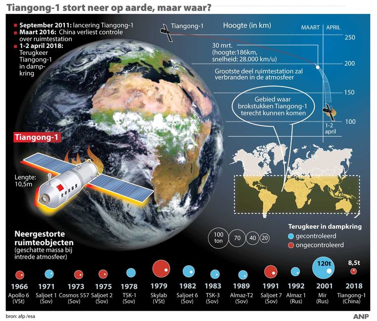
Zo groot als een bus en 9 ton zwaar, maar waar en wanneer stort Chinees ruimtestation dit weekend neer op aarde? - HLN.be
Zo groot als een bus en 9 ton zwaar, maar waar en wanneer stort Chinees ruimtestation dit weekend neer op aarde? - HLN.be
Infografieken Nederland
WETENSCHAP Dit weekend zal het Chinese ruimtestation Tiangong-1, dat ongeveer zo groot is als een bus, met een rotvaart op aarde neerstorten. De kans dat je een van de brokstukken op je hoofd krijgt, is wel bijzonder klein: er is maar één kans op een biljoen.
“Wanneer en waar het station zal neerkomen, is niet precies te voorspellen”, weet Bart Hendrickx, die als ruimtevaartspecialist verbonden is aan Volkssterrenwacht Urania. “De laatste voorspelling is dat het zondag zal zijn. Zeker is dat de brokstukken ergens tussen 43° noorderbreedte en 43° zuiderbreedte zullen neervallen. Wij lopen dus helemaal geen gevaar.” Het gebied waar stukjes van het ruimtestation kunnen terechtkomen, strekt zich ongeveer uit van het midden van Italië tot onder Australië. Vrij groot, inderdaad. “Maar”, zegt Hendrickx, “Vergeet ook niet dat 70% van de aarde uit water bestaat. De kans dat je getroffen wordt door een blikseminslag is vele keren groter.”
Het ruimtestation, dat in 2011 gelanceerd werd en waarmee de Chinezen al in 2016 het contact verloren, is zo groot als een autobus. “Het weegt ongeveer 9 ton en is 10 meter lang. Maar er zijn al veel grotere objecten naar beneden gekomen: het Amerikaanse ruimtestation Skylab, dat in 1979 neerstortte, woog 80 ton of met andere woorden tien keer meer. Dat Tiangong-1 - wat Chinees is voor ‘Hemels Paleis’ - ongecontroleerd naar beneden zal komen, is geen ideaal scenario, maar ook dat is in het verleden nog gebeurd.”
Tiangong-1 zal neerstorten met een snelheid van 27.000 kilometer per uur, wat volgens Hendrickx véél trager is dan een meteoriet. Bovendien zullen de meeste brokstukken opbranden bij contact met de dampkring. “Als je toch nog een stuk vindt van het ruimtestation, kan je er beter afblijven: de temperatuur zal hoog zijn en in de tank kan ook altijd wat van de giftige brandstof achtergebleven zijn.”
REUTERSEen schaalmodel van het bewuste ruimtestation.
I suspect that a lot of UFO activity in the latter part of the 1940s in the United States had more to do with classified, military experimentation than it did with aliens. There is, however, no doubt that there was a significant number of UFO reports from that period which are not so easy to dismiss. In many such cases, UFOs intruded upon sensitive military/government airspace in the United States. In the latter part of the 1940s – and particularly so in 1948 – a curious phenomenon was repeatedly seen in the skies over New Mexico. They were strange, glowing balls of green light that seemed to take a disturbing amount of interest in the many defense, atomic, and military establishments that existed in the state at the time. A program designed to investigate the reports, which was given the name of Project Twinkle, was soon put into place.
Consider the following, written by Lt. Col. Doyle Rees of the Air Force’s Office of Special Investigations (AFOSI) at Kirtland Air Force Base, New Mexico, on 25 May 1950. It was for the specific attention of Brigadier General Joseph F. Carroll, Director of Special Investigations, USAF:
“In a liaison meeting with other military and government intelligence and investigative agencies in December 1948, it was determined that the frequency of unexplained aerial phenomena in the New Mexico area was such that an organized plan of reporting these observations should be undertaken. The organization and physical location of units of this District were most suitable for collecting these data, therefore, since December 1948, this District has assumed the responsibility for collecting and reporting basic information with respect to aerial phenomena occurring in this general area.”
Los Alamos
And a large body of that so-called “basic information” was profound, to say the very least. A Federal Bureau of Investigation (FBI) memorandum dated 31 January 1949 and titled Protection of Vital Installations, reveals the following: “During the past two months various sightings of unexplained phenomena have been reported in the vicinity ofthe Atomic Energy Commission Installation at Los Alamos, New Mexico,where these phenomena now appear to be concentrated[emphasis mine]. During December 1948 on the 5th, 6th, 7th, 8th, 11th, 13th, 14th, 20th, and 28th sightings of unexplained aerial phenomena were made near Los Alamos by Special Agents of the Office of Special Investigations; Airline Pilots; Military Pilots; Los Alamos Security Inspectors; and private citizens. On January 6, 1949, another similar object was sighted in the same area.” Moving on…
It’s intriguing to note that the atomic research facility at Oak Ridge, Tennessee was also a magnet for countless close encounters with UFOs – in the late 1940s and the early 1950s. Consider, for example, the following official report generated on 2 March 1950 by the Atomic Energy Commission (AEC):
“There is a radar station near Knoxville which has been in operation about 3 weeks. This radar station is being operated by station WROL of Knoxville. On 1 March at 2135 hours the station picked up an object 340 degrees and 18 miles from Knoxville, altitude 40,000 feet. Direction and distance put the object directly over [emphasis mine] Oak Ridge. AEC Security Division Chief at Oak Ridge checked with Smyrna Air Base, Nashville which reported it had no flight plan for any plane in that vicinity and altitude. On 2 March at 1105 station picked up object at 335 degrees and 18 miles from Knoxville, altitude 40,000 feet. AEC Security Division Chief checked with Smyrna Air Base with negative results.”
On the following day, 3 March, 1950, a further report surfaced: “At 2130 hours on 2nd March, radar station picked up two objects 310 degrees, altitude 80,000 feet, approximately 18 miles from Knoxville in general direction of Oak Ridge, moving in circular motion but in opposite directions.” And Oak Ridge continued to be a target for the mysterious intruders. An FBI teletype of 13 October 1950 refers to the detection of a veritable squadron of unknown objects tracked over the Oak Ridge installation at 11.25 p.m. on 12 October: “USAF radar installation at Knoxville picked up indications of eleven objects and perhaps more traveling acrosscontrolled areaof Atomic Energy installation at Oak Ridge [emphasis mine]…operators are experienced reliable personnel and radar set is in perfect operating condition.”
Oak Ridge
Perhaps most significant of all, is the following FBI document that offers the significant thoughts and conclusions of Oak Ridge personnel with respect to the UFO invasions in sensitive airspace:
“The opinions of the Security Division, AEC Oak Ridge; Security Branch, NEPA [Nuclear Energy for Propulsion of Aircraft project] Division, Oak Ridge; AEC Security Patrol, Oak Ridge; FBI Knoxville; Air Force Radar and Fighter Squadrons, Knoxville; and the OSI, Konxville, Tennessee, fail to evolve an adequate explanation for OBJECTS SIGHTED OVER OAK RIDGE, TENNESSEE; however, the possibilities of practical jokers, mass hysteria, balloons of any description, flights of birds, falling leaves, insect swarms, peculiar weather conditions, reflections, flying kites, objects thrown from the ground, windblown objects, insanity, and many other natural happenings have been rejected because of the simultaneous witnessing of the objects with the reported radar sightings; because of the reliability of the witnesses; because of the detailed, similar description of the objects seen by different persons…”
Note that in the above-paragraph the words “objects sighted over Oak Ridge” were all typed in capital letters, further demonstrating the concern over such cases.
Not exactly outright invasions, but pretty damn close.
This is no April Fool’s joke. The dead-and-descending-out-of-control Chinese space station Tiangong-1 is predicted to crash somewhere on Earth on April 1st, plus-or-minus a day or two. This was probably not a good time for the recent U.S. government actions against China because its government and space agency hasn’t been too open about when or where the space debris might make its flaming landfall. The Chinese media site Global Times got an official to speak but only on the condition of anonymity and he said it’s impossible to pinpoint the exact reentry location now (March 29). In fact, it won’t be known until just two hours before it starts to fall. Thanks, China!
I thought you said I had two hours!
Fortunately, scientists at the International Centre for Radio Astronomy Research have no fear for their lives (from their government, at least) and are willing to share some signs that the end of the Heavenly Palace (the meaning of Tiangong) is near.
“[You will see a] series of fireballs streaking across the sky.”
Fireballs? That’s it? No explosions? Horns? Apocalyptic voices from the sky? That helpful hint is from Markus Dolensky, of the International Centre for Radio Astronomy Research, a joint venture between Curtin University and the University of Western Australia. That’s not much from some of the world’s leading researchers in radio astronomy who are building the International Square Kilometre Array — the world’s biggest ground-based telescope array.
Maybe an astrophysics researcher is a better person to ask than an astronomer. The Daily Star checked with Alan Duffy, an astrophysics researcher at Swinburne University of Technology in Australia, and here’s what he had to say.
“The international community doesn’t know what the craft is made of, and that makes estimating the danger more challenging, as hardened fuel containers could reach the ground while lightweight panels won’t.”
So, we need to be looking for fireballs of space station fuel, which we already know contains hydrazine – a highly-toxic propellant used by China in rocket fuels. The U.S. Environmental Protection Agency says short-term exposure to high levels of hydrazine – burning or otherwise — may cause irritation of the eyes, nose, and throat, dizziness, headache, nausea, pulmonary edema, seizures, coma and cancer. Good thing we’ll be able to see it, right? Well, only if it’s burning. Otherwise, it’s a colorless liquid with an ammonia smell. You know … like ammonia.
Is there ANY chance this is an April Fool’s joke?
If we survive, the next time a space station crashes to Earth it won’t be so bad, according to Kenneth Chang of The New York Times.
“Indeed, space agencies like the E.S.A. are using Tiangong-1 as a learning exercise to compare their prediction models.”
April Fool’s Day was so much more fun when the biggest story was Taco Bell buying the Liberty Bell and renaming it the Taco Liberty Bell.
Let’s hope we have a similarly good laugh about Tiangong-1 on April 2nd.
Today, I’m going to tell you the intriguing story of Project Horizon. It fell under the auspices of the U.S. Army, and had its origins in the latter part of the 1950s. The plan was to take the first steps towards constructing an installation on the surface of the Moon by the mid-1960s. The goal was for the base to be armed with nuclear weapons and which could be used to decimate the Soviet Union if the United States was hit by a sneak attack and the infrastructure of the nation was significantly destroyed.
After much preliminary discussion, it was in late March 1959 that the ambitious program was finally put into place. Overseeing many of the plans to create the secret base was Lieutenant General Arthur G. Trudeau. At the time, Trudeau was the Army’s Chief of Research and Development. Now-declassified files on Project Horizon demonstrate that Trudeau and his team estimated it would cost approximately $6 Billion to design, build and fully equip a base on the Moon. In a document titled “Project Horizon: A U.S. Army Study for the Establishment of a Lunar Military Post,” Trudeau wrote the following words:
“There is a requirement for a manned military outpost on the Moon. The lunar outpost is required to develop and protect potential United States interests on the Moon; to develop techniques in Moon-based surveillance of the Earth and space, in communications relay, and in operations on the surface of the Moon, for further exploration into space and for military operations on the Moon if required; and to support scientific investigations on the Moon.”
The program was indeed a grand one, a far-reaching operation designed to ensure that the control of outer space would fall into the hands of the United States and and specifically not into the hands of the Russians. In the wake of the publication of Trudeau’s report, plans were quickly initiated. One of those who was brought into the program was Wernher Von Braun – a scientist, and someone who had been a member of Germany’s Nazi Party and a member of the SS. He was an expert in the field of rocketry and someone who, after the Second World War, was brought to the United States under cover of a classified and controversial program called Operation Paperclip. Von Braun chose to assign an engineer named Heinz-Hermann Koelle to oversee Project Horizon. Koelle fought for the Nazis during the Second World War. Evidently, the Nazi-based backgrounds of the pair didn’t bother those working on Project Horizon. It certainly should have, though. But, I digress.
The initial goal was for the Moon base to be relatively small, which made a great deal of sense. After all, we’re talking about entirely new territory and revolutionary technology. So, the plan was to slowly make the base bigger and bigger as time went on. But, initially, it would be something akin to a North Pole outpost, with a staff of around just one to two dozen. Using the base as a strategic military facility, as well as a place in which the mysteries of the Moon and the Solar System could be carefully and secretly studied, ensured that America would have a significant lead over the Soviets – who were clearly a major threat at the time.
In fact, on this issue of a potential Soviet threat, the Project Horizon team gave serious and deep consideration to the possibility that the Russians might very well try and destroy the base – possibly even with Russian cosmonauts invading the base and armed to the teeth with high-tech weaponry. On the scientific side of things, a great deal of research was focused on ensuring that the base would have a plentiful supply of water and oxygen – which, of course, none could live without. Plans were initiated to have vast shuttle craft send endless supplies of water and food to the base – that is, until the staff became completely self-sufficient, which was seen as completely feasible. In theory, at least.
As for how and where, exactly, the base would be constructed, Project Horizon’s scientists were of the opinion that the best option would be to build it in an already-existing, natural crater. Such was the enthusiasm for the program, an estimation was made that the initial construction of the base could begin in 1965 – which was four years before Neil Armstrong set foot on the surface of the Moon. Project workers suggested that it might be wise to construct significant parts of the base underground, chiefly to protect it from not just the Soviets, but from natural space debris too, such a fragments of meteorites.
Although Project Horizon was seen as a major (and secret) program of the military, it was ultimately deemed to be way too far ahead of itself. The military concluded that the plans for the creation of Project Horizon to begin in 1965 were overly ambitious in the extreme and the program was cancelled. What was once on the horizon was finally no more.
“We are researchers at Johns Hopkins University working on a research study regarding the experiences of people who have had encounters with seemingly autonomous beings or entities after taking DMT.”
Kids, don’t try this at home (DMT is illegal in most countries), but if you already have seen aliens while on DMT, researchers at Johns Hopkins University School of Medicine led by Roland Griffiths, Ph.D. would like for you to fill out an anonymous survey on your experiences and visions. You must be at least 18 years old, fluent in English and have taken a “breakthrough” dose of N,N-DMT before the alien experience.
“During your encounter, was there any communication (e.g., a one-way or two-way exchange of information) between you and the being or entity?”
DMT or N,N-DMT is N,N-Dimethyltryptamine, the tryptamine molecule often consumed as a psychedelic drug in ayahuasca, particularly in Amazon cultures, and sometimes referred to as the ‘spirit molecule’ because of the mystical experiences and dramatic hallucinations it causes. During the 1960s it was called the “businessman’s trip” because, when smoked or injected, the hallucinations arrive almost immediately and lasted just a short time – 5 to 15 minutes. That’s not the case when it is consumed in an ayahuasca beverage prepared by a South American shaman. Growing in popularity, these experiences can last up to 6 hours and the intense physical trauma (tremors, diarrhea, hyperthermia, sweating, muscle spasms) are considered to be a small price to pay for them. It’s this type of experience that most likely generates the alien visions Dr. Griffiths is looking for.
“Did the being or entity seem to have an emotional response to it’s encounter with the you (this could include being consciously unemotional or unreactive)?”
Stories of encounters with beings or entities while under the influence of DMT are not uncommon. A 2012 article in Psychology Today reveals that Rick Strassman, a leading expert on hallucinogenic substances and the coiner of the term “spirit molecule,” found that about half of the 60 volunteers who took DMT in one of his studies in the early 1990s entered “freestanding, independent levels of existence” that were inhabited by what volunteers described as intelligent “beings”, “entities”, “aliens”, “guides”, and “helpers” that appeared as “clowns, reptiles, mantises, bees, spiders, cacti, and stick figures.” According to the article, seeing beings and entities of these types seems to be unique to DMT — Strassman was unable to find anything similar in research reports on other psychedelic drugs.
“Did you ascertain a message, task, mission, purpose, or insight from your encounter with the being or entity (this could have been directly imparted to you or attained through your understanding of the nature of the being, etc.)?”
The questionnaire is extensive and obviously focused on determining if there is a link between DMT and actual alien encounters or the illusion of them.
Here are some other questions asked:
Did you acquire any predictions about the future through your encounter with the being or entity? Do you think that the being or entity continued to exist after your encounter? Was the being or entity all-knowing? How clear are your memories of the experience? Overall, from your current perspective, which of the following best describes where you think the entity existed? Do you believe that the experience and your contemplation of that experience has altered your fundamental conception of reality?
If you’re interested and qualified and would like to participate in the anonymous research, the questionnaire is located here.
Do you believe they will find a link between DMY and aliens – real or imagined? Do you already KNOW there’s one?
If it were to be proven that DMT was a gateway drug to an ET gateway, would you consider taking it?
0
1
2
3
4
5
- Gemiddelde waardering: 0/5 - (0 Stemmen) Categorie:ALIEN LIFE, UFO- CRASHES, ABDUCTIONS, MEN IN BLACK, ed ( FR. , NL; E )
More Strange Waves Of Colored Light Fill The Sky
More Strange Waves Of Colored Light Fill The Sky
Strange glows as what are being described as "strange waves of colored light" in the night sky a couple of hours or so after Sunset has been observed again.
Observers from Pennsylvania as well as observers from Athens Gorgia describe the colored sky waves of light that do resemble auroras,
They witnessed the strange glow off and on for 40 minutes or so in Georgia looking to the NW. All of the observers had never seen anything like this before!
UFO Over River In Minnesota Near Small Town, Photos, UFO Sighting New
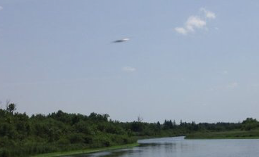
UFO Over River In Minnesota Near Small Town, Photos, UFO Sighting News.
Date of sighting: 7-4-2011 Location of sighting: Nimrod, Minnesota, USA Source: MUFON #91099 This UFO was seen back in 2011, but reported today. The photo was taken in the city of Nimrod which has a sparse population of just around 67. UFOs are known to be frequently seen around areas where there are less humans. The UFO is a disk viewed from the side. The suns reflection off its metallic surface gives it away. Scott C. Waring
Eyewitness states:
Picture was taken on a 4th of July camping trip in 2011. Noticed the object when going through old photos this week.
An illustration of India's Chandrayaan-2 lunar spacecraft.
Credit: ISRO
Chandrayaan-2 is India's planned second mission to the moon, which is expected to launch in 2018. It is a follow-up mission from the Chandrayaan-1 mission that assisted in confirming the presence of water/hydroxyl on the moon in 2009. Chandrayaan-2 will launch from the Satish Dhawan Space Center in Sriharikota, India, aboard a Geosynchronous Satellite Launch Vehicle (GSLV) rocket.
According to the Indian Space Research Organization (ISRO), the new mission will consist of an orbiter, a lander and a rover. The orbiter will perform mapping from an altitude of 100 kilometers (62 miles), while the lander will make a soft landing on the surface and send out the rover.
Development and science
Initially, ISRO planned to partner with Russia to perform Chandrayaan-2. The two agencies signed an agreement in 2007 to launch the orbiter and lander in 2013. Russia later pulled out of the agreement, however, according to a news report from The Hindu. The Russian lander's construction was delayed after the December 2011 failure of Roscosmos' Phobos-Grunt mission to the Martian moon of Phobos, the report stated.
Russia subsequently pulled out of Chandrayaan-2 altogether, citing financial issues. Some reports stated that NASA and the European Space Agency were interested in participating, but ISRO proceeded with the mission on its own.
The Chandrayaan-2 orbiter will circle the moon and provide information about its surface, ISRO stated. "The payloads will collect scientific information on lunar topography, mineralogy, elemental abundance, lunar exosphere and signatures of hydroxyl and water-ice," ISRO said on its website. The mission will also send a small, 20-kilogram (44 lbs.), six-wheeled rover to the surface; the rover will move semi-autonomously, examining the lunar regolith's composition.
Terrain Mapping Camera 2 (TMC-2), which will map the lunar surface in three dimensions using two on-board cameras. A predecessor instrument called TMC flew on Chandrayaan-1.
Collimated Large Array Soft X-ray Spectrometer (CLASS), which will map the abundance of minerals on the surface. A predecessor instrument called CIXS (sometimes written as C1XS) flew on Chandrayaan-1.
Solar X-ray Monitor (XSM), which looks at emissions of solar X-rays.
Chandra's Atmospheric Composition Explorer (ChACE-2), which is a neutral mass spectrometer. A predecessor instrument called CHACE flew on Chandrayaan-1's Moon Impact Probe.
Synthetic Aperture Radar (SAR), which will map the surface in radio waves. Some of its design is based on Chandrayaan-1's MiniSAR.
Imaging Infra-Red Spectrometer (IIRS), which will measure the abundance of water/hydroxl on the surface.
Orbiter High Resolution Camera (OHRC) to examine the surface, particularly the landing site of the lander and rover.
The lander's instruments include:
Instrument for Lunar Seismic Activity (ILSA), to look for moonquakes.
Chandra's Surface Thermophysical Experiment (ChaSTE), to examine the surface's thermal properties.
Radio Anatomy of Moon Bound Hypersensitive ionosphere and Atmosphere (RAMBHA-Langmuir Probe), to look at plasma density on the surface.
The rover will carry two science instruments to look at the composition of the surface: the Laser-Induced Breakdown Spectroscope (LIBS) and the Alpha Particle X-Ray Spectrometer (APXS).
Landing near the pole
Chandrayaan-2's lander and rover are targeted for a location about 600 kilometers (375 miles) from the south pole, which would be the first time any mission touched down so far from the equator, according to a January 2018 article in Science magazine. ISRO plans to use the experience for more challenging missions in the future, such as touching down on an asteroid or Mars, or sending a spacecraft to Venus, IRSO chair Kailasavadivoo Sivan said in the article.
The lander is expected to last about one lunar day, or 14 Earth-days; it is unclear if it will revive after falling into the darkness of a lunar night. Its measurements of moonquakes would provide more data after the information collected by the human Apollo missions in the 1960s and 1970s, according to Science. Meanwhile, the terrain explored by the rover is of great scientific interest due to the rocks' old age; at 4 billion years old, scientists say the zone likely is composed of an ancient magma ocean.
While NASA is not directly participating in this mission, the measurements taken by Chandrayaan-2 could be a help for future lunar missions. That is because in late 2017, the agency was tasked by the Trump administration to return humans to the moon in the coming years.
Telescope Spots Doomed Chinese Space Station (Photo)
Telescope Spots Doomed Chinese Space Station (Photo)
By Hanneke Weitering, Space.com Staff Writer
Astronomers with the Virtual Telescope Project captured this view of Tiangong-1 on March 28, 2018, using the Tenagra III "Pearl" telescope at the Tenagra Observatories in Arizona. The image is a single, 2-second exposure captured at 8:35 a.m. EDT (1235 GMT).
With just a few days left before China's Tiangong-1 space station is expected to come crashing down on Earth, astronomers captured this incredible view of the derelict craft zooming through space.
The Virtual Telescope Project in Italy, together with the Tenagra Observatory in Arizona, used a robotically controlled telescope to provide live views of the space station during a webcast this morning (March 28). In this view from the webcast, Tiangong-1 is gleaming as the sun reflects off the spacecraft's surface and faint star trails streak across the background.
Capturing the out-of-control space station "was an extremely difficult task" due to the craft's speed, Gianluca Masi of the Virtual Telescope Project said in a statement. Tiangong-1 was moving at about 18 degrees per minute (for comparison, your clenched fist held at arm's length is about 10 degrees wide). According to the satellite-tracking website satview.org, the station's speed was approximately 17,400 mph (28,000 km/h). [China's Space Station Crash: Everything You Need to Know]
"Our telescope honestly did out-of-this world work to track such a target," Masi told Space.com in an email. Tracking an object that swift "is something which very few observatories can do," Masi said, but the telescopes available at the Virtual Telescope Project and Tenagra Observatory were equipped for the challenge. The Paramount ME robotic mount installed on these telescopes is "the best hardware of this kind available in the world," the Virtual Telescope Project's website states.
At the time the image was captured, Tiangong-1 was orbiting at an altitude of about 137 miles (220 kilometers). That's around half as high as the International Space Station, which flies at an average altitude of 248 miles (400 km) above the Earth.
Tiangong-1's orbit is slowly decaying, and the station is expected to re-enter Earth's atmosphere sometime between March 31 and April 1, according to the European Space Agency (ESA). While most of the station will burn up in the atmosphere, some of its hardier components will likely survive the inferno and crash into Earth, experts predict. But there's no need to hunker down all weekend; ESA officials estimate that the odds of being stuck by a piece of Tiangong-1 are less than 1 in 300 trillion, and the station will most likely fall in the ocean.
Email Hanneke Weitering at hweitering@space.com, or follow her @hannekescience.
Filer’s Files #12 – 2018 Spaceships Surround Earth - PART I
John Lenard Walson captured this video of a ship in space
Filer’s Files #12 – 2018 Spaceships Surround Earth - PART I
In special reports, this week’s files cover: Admiral Lord Hill-Norton UFOs Are Real, Is Trump playing the Reagan Space Defense card? Space Objects Photographed, President Reagan’s Diary, Air Force to Defend Space, Thom Reed UFO Monument Park, Letter About Spaceship on NASA TV, and Thom Reed UFO Monument Park.
In special reports, this week’s files cover: Unidentified Aerial Phenomena sightings were reported over Alabama, Arizona, California, Colorado, Florida, Michigan, New Jersey, Pennsylvania, and Texas..
Unidentified Aerial Phenomena sightings were reported over Austria, Belgium, Canada, France, Iceland, Northern Ireland and England in the United Kingdom.
The Filer Research Institute feels the scientific study of UFOs is for the benefit of humankind and is an important endeavor. The US Air Force investigated UFOs publicly for more than twenty years under Project Blue Book; and I continue this advanced research. I believe the God of the universe has spread life throughout the cosmos and UFO’s are visiting us in ever-increasing numbers.
Forward these files to your friends and neighbors.
Special Reports
Admiral Lord Hill-Norton UFOs Are Real
Admiral of the Fleet Lord Hill-Norton wrote, “The evidence that there are objects which have been seen in our atmosphere, and even on terra firma, that cannot be accounted for either as man-made objects or as any physical force or effect known to our scientists, seems to me to be overwhelming.
A very large number of sightings have been vouched for by persons whose credentials seem to me unimpeachable. It is striking that so many have been trained observers, such as police officers and airline or military pilots. Their observations have in many instances… been supported either by technical means such as radar or, even more convincingly, by… interference with electrical apparatus of one sort or another….
Lord Hill-Norton, Chief of Defense Staff, Ministry of Defense, Great Britain, 1973; He became Chairman of the NATO Military Committee in 1974, remaining in that post until his retirement in 1977. Quoted from his foreword to “Above Top Secret” by Timothy Good, Morrow & Co’s Quill Books, 1988. _
Is Trump playing the Reagan Space Defense card? To what end?
Clarkson writes, “In a somewhat surreal scenario, President Donald J. Trump last week remarked on the idea of a “Space Force,” something he’d clearly been briefed on at some point. After telling an audience at Marine Corps Air Station Miramar that “space is a war fighting domain,” Trump tossed off the idea “that “we may even have a space force.” Also the Air Force announced that the 200 man National Space Defense Center will operate 24 hours seven days a week to protect US satellites. It appears offensive forces would be needed conduct combat operations to protect the satellites. The following stories may show what in space.
Space Objects Photographed
Objects photographed in space several hundred miles above Earth include the ISS and are likely either human space craft such as Solar Warden or extraterrestrial craft. This is an alleged top secret aircraft or “space vehicle” which has some quite incredible abilities. The source of the information is from a former Area 51 employee who started making public disclosures in 1998 after retiring from his job as an avionics engineer. Huge Objects are regularly photographed in space above the Earth by John Leonard Walson.
In 2007, a John Lenard Walson invented a new gadget capable of extending his amateur telescope resolution. The new way to extend the capabilities of his eight inch Meade telescope to shoot his videos has enabled him to achieve optical resolutions – at almost the diffraction limit – not commonly achievable. The discovery was first publicized by Jeff Rense at “Mystery Space Machines Above. Black Ops, Star Wars Or ET? Or All Of The Above?” in December 2007.
Maybe some of Walson’s images are sensitive, secret US or Russian military Star Wars machines. On March 23, 1983, President Reagan proposed the creation of the Strategic Defense Initiative (SDI), an ambitious project that would construct a space-based anti-missile system. The SDI was intended to defend the United States from attack from Soviet ICBMs . Maybe these ‘secret’ weapon platforms in space were actually deployed, which the US military has been rumored to have for at least 20 years. It’s clear the major space powers are far more heavily-invested in space than they will admit. Of course the major powers know what each other has in space. If they have been there for thirty years they are getting obsolete.
As a reflection of that reality, there has been a lot of recent talk about ‘anti-satellite’ weapons ‘needed’ by competing countries to ‘protect themselves.’ Most recently, China has been discussing its anti-sat programsand even threatened to destroy or disable all GPS satellites which over-fly Chinese territory. It’s anyone’s guess how many billions the US military and government have poured into lack ops. Walson’s video images and sounds may well show some of these advanced machines. So, what you see could be spacecrafts, and maybe even space ships, parked or stationed in orbit above the Earth. It is also within the realm of possibility some of these items might be products of non-human intelligence.
These are not just satellites and space junk? We are seeing large and very sophisticated spacecraft that rockets are unlikely able to put in orbit. Another propulsion system would be needed suggesting alien technology. Walson received the following comment about one of the videos:
“My congratulations on your superb astrophotography. MIT Lincoln Laboratory is the group which has built some of the things you are seeing. Much of what they do is what used to be the Star Wars project, which no doubt involves some of your objects.” MIT Lincoln Laboratory is federally funded and was sponsored by the Missile Defense Agency, and Defense Advanced Research Projects Agency. It is known they examined crashed UFOs held at Wright Patterson AFB and likely back engineered these craft.
Here is what the International Space Station (ISS) looks like through the telescope and video camera lens of astro-photographer Walson. The dimensions of the ISS research is 356 feet (109 meters) by 240 feet (73 meters), or slightly larger than a football field.. It is the International Space Station… the ISS… take a look… Keeping that image and it’s relative size appearance in mind, now ask yourself, what is THIS? It looks at least 500 feet long or almost two footbsll fields.
So far his claims and disclosures have not been given the coverage which they undoubtedly deserve. He has hundreds of official USAF documents to back up his claims. If he is correct, as the evidence suggests, then the U.S. does have a secret space fleet consisting probably of dozens of 600 feet wide flying triangles, which can go to and from space with ease, travel at Mac 9 +in any direction. For over ten years John Walson has taken up close images of celestial craft in orbit around the Earth John writes, ‘I have so much more that I have not told anyone yet, I have very specially made UV spectrum telescopes and UV real cameras with very amazing footage. I would really like to share my telescope inventions in a TV show to use my equipment to solve famous historical Ufo or Sky space based events.And here is the actual video footage from which the above freeze frame is taken followed by some footage of something.
I’m saving to be able to bring my telescope to a dark area, no atmospheric distractions. I want to give you clear beautiful high quality video, and evidence from a remote location without freezing in the process, and without interference.
Walson claims he discovered that many “stars” in the sky are in fact HUGE structured objects – giant machines are “fixed” in the sky. With this new-found ability, he has proceeded to videotape, night and day, many strange and heretofore unseen objects in earth orbit. Walson claims that he can record them in plain daylight at noon and that he can record sounds from the machines in space. The resulting astro-photographic video footage has revealed a raft of machines, hardware, satellites, spacecraft and likely alien space ships which otherwise appear as ‘stars’… if they appear at all. These discs are seen hovering over thunderstorms appearing to collect lightning strikes perhaps to collect electrical energy.
These discs are seen hovering over thunderstorms appearing to collect lightning strikes perhaps to collect electrical energy.
Note: John has given Filer’s Files permission to show his excellent copyrighted images. Below is a possible space ship taken by John that President Reagan refers to.
In The Diary entry for Tuesday, June 11, 1985 (page 334) Reagan wrote:
Lunch with 5 top space scientists. “It was fascinating. Space truly is the last frontier and some of the developments there in astronomy etc. are like science fiction, except they are real. I learned that our shuttle capacity is such that we could orbit 300 people.”
Note: This would indicate a space ship at least as large as a 747 aircraft that is 250 feet long. It is likely much bigger to accommodate sleeping quarters, kitchen facilities, bathrooms, storage radar and weapons.The now grounded Space Shuttle held a maximum of eight people and even if all five took off fully loaded it would be impossible to put 300 astronauts in orbit. Apparently President Reagan revealed the existence of a highly classified space program that could accommodate hundreds of astronauts in orbit that would to be at least 500 feet long?
British hacker Gary McKinnon first hacked into NASA’s Johnson Space Center: He found a high definition picture of a large cigar shaped object over the northern hemisphere.
McKinnon later hacked into classified files of U.S. Space Command incorporated into Strategic Command on October 1, 2002. He found a list of officers’ names. Under the heading ‘Non-Terrestrial Officers. McKinnon stated that what he found was a list of fleet-to-fleet transfers’ ‘fleet-to-fleet transfers, and a list of ship names’. He said that after looking them up. He realized that they were not US Navy ships. What he saw made him believe that NASA was keeping a secret about an off planet space fleet. Gary McKinnon claims to have stumbled upon a secret list of up to ten space warships after breaking into NASA computer databases. A couple ships were named the USSS Curtis Lemay and USSS Roscoe Hillenkoetter. He claims it points to a secret American space program.
Air Force to Defend Space
Secretary of the Air Force Heather Wilson testified before the U.S. House of Representatives Armed Services Committee about the Air Force’s fiscal year 2019 budget on March 20, 2018, in Washington, D.C. “The Air Force budget for FY19 aligns with the National Defense Strategy,” said Wilson. “In our budget, there are really two bold moves and one continuing theme. The first bold move is the acceleration of a defendable space. The Air Force, Wilson said, needs to be able to deter, defend and prevail against anyone who seeks to deny the nation’s ability to operate freely in space.
“The United States of America is the best in the world at space and our adversaries know it. In any future conflict we expect that they will seek to deny us the use of space.
So what we’re doing in this budget is accelerating our ability to defend our assets on orbit,” she said.The Air Force operates 76 satellites, 30 of which are GPS and another 25 are communication satellites. According to Wilson, the service is investing in jam-resistant satellite technology for both communications and GPS capabilities. Seth Moulton (D-Mass.) pointed to “China’s development of anti-satellite weapons and how they now claim to be able to take out the majority of our satellites within 28 hours, 48 hours rather at the beginning of a conflict.”
“The second bold move in this budget is the shift to multi-domain operations and that’s most visible in the way the Air Force plans to do command and control,” Wilson said. “There is also one continuing effort in our budget and that is to keep improving readiness to win any fight any time, that’s what you expect of your Air Force.” And the defense budget sequester “did more damage to the United States Air Force and our ability to defend the nation than anything our advisories have done in the last 10 years — we did it to ourselves,” Wilson said. “We cut 30,000 people out of the Air Force, reduced [the force] by 10 fighter squadrons, and [reduced] weapons systems sustainment,” she added. Problems with pilot retention can be tied directly back to sequester, Wilson said.
X-37b Space ship at Space Council Meeting.
Secretary Heather Wilson at the recent Space Council seemed to commit the US to more “offensive” weapons for space war “We have to be able to do something if things are going wrong,” Wilson said at the Center for Strategic and International Studies. “Offensive” weapons will be needed. “We need the ability to create effects, defend ourselves and deter actions,” she said. Her comments are the latest in what is clearly a serious increase in attention — if not funding — for offensive space weapons.
Note: Who in space is are potential enemy?
F-l8s Intercept UFO
Commander David Fravor and Lt. Cmdr. Jim Slaight were on a routine training mission 100 miles out into the Pacific when the radio in each of their F/A-18F Super Hornets crackled: An operations officer aboard the U.S.S. Princeton, a Navy cruiser, wanted to know if they were carrying weapons.
“Two CATM-9s,” Commander Fravor replied, referring to dummy missiles that could not be fired. He had not been expecting any hostile exchanges off the coast of San Diego that November afternoon in 2004.
Commander Fravor, in a recent interview with The New York Times, recalled what happened next. Some of it is captured in a video made public by officials with a Pentagon program that investigated U.F.O.s.
“Well, we’ve got a real-world vector for you,” the radio operator said, according to Commander Fravor. For two weeks, the operator said, the Princeton had been tracking mysterious aircraft. The objects appeared suddenly at 80,000 feet, and then hurtled toward the sea, eventually stopping at 20,000 feet and hovering. Then they either dropped out of radar range.
The radio operator instructed Commander Fravor and Commander Slaight, who has given a similar account, to investigate.
The two fighter planes headed toward the objects. The Princeton alerted them as they closed in, but when they arrived at “merge plot” with the object — naval aviation parlance for being so close that the Princeton could not tell which were the objects and which were the fighter jets — neither Commander Fravor nor Commander Slaight could see anything at first. There was nothing on their radars, either.
Then, Commander Fravor looked down to the sea. It was calm that day, but the waves were breaking over something that was just below the surface. Whatever it was, it was big enough to cause the sea to churn.
Hovering 50 feet above the churn was an aircraft of some kind — whitish — that was around 40 feet long and oval in shape. The craft was jumping around erratically, staying over the wave disturbance but not moving in any specific direction, Commander Fravor said. The disturbance looked like frothy waves and foam, as if the water were boiling.
A video shows a 2004 encounter near San Diego between two Navy F/A-18F fighter jets and an unknown object. It was released by the Defense Department’s Advanced Aerospace Threat Identification Program.
By Courtesy of U.S. DEPARTMENT OF DEFENSE on Publish Date December 16, 2017. Photo by U.S Department of Defense.
Commander Fravor began a circular descent to get a closer look, but as he got nearer the object began ascending toward him. It was almost as if it were coming to meet him halfway, he said.
Commander Fravor abandoned his slow circular descent and headed straight for the object.
But then the object peeled away. “It accelerated like nothing I’ve ever seen,” he said in the interview. He was, he said, “pretty weird.”
Letter About Spaceship on NASA TV
Hi Major I hope you’re doing well. this is Mike Forte’ X State section Director Tampa Bay. I was watching the Earth Turn on the nasa Channel. I often do it beats the news. Its peaceful and breath taking. of course I’m always looking for a Star Wars Weapon to shoot up and try to nail .something as they did back in the 90s on film. I never see anything too weird until 7 30 PM, I see the nasa logo always on the right but for once it had a counterpart on the left side bigger and straight.. At this point I’m focused because as I watch I realize its moving and turning away from earth. I’m trying to figure out what kind of new graphic I was seeing. I would it was about 10 or 20 miles long. angular from rear to front darkish blue. It had to be 10 to 15 miles above the earth. As it turned away I could see the rear. This is no logo with a wide oblong like rectangular shape defined as it floated further away, getting smaller. I thought could be ours but no way unless the many years of stories we’ve heard are out of wack about what we do possess. Clean lines no windows or shapes definable..I have been involved in cases with these type ships as part of the criteria. Without question I watched one and dam near went blind watching it.
I did what I could to draw a pic someone else had to see it too. If someone didn’t believe in these ships this might change their mind from the sheer size alone. I’ve seen a lot of real stuff in my 45 years of investigating but NASA gets the award for slipping this one in. I can only wonder what the guys in the Space Shuttle saw and filmed. Thanks to Mike.
“Thom Reed UFO Monument Park”
Kerry McClure writes, There’s a new UFO destination in, New England – The Thom Reed UFO Monument Park, a waterfront park that now stands at the location of the 1st UFO incident officially inducted into the United States as historically true. The park, which represents modern-day history and a progressive effort to observe such events, is free to the public and located at the precise location of the incident.
The monument is nestled in front of a replica of the old covered bridge – the bridge the Reeds drove through that September night, just seconds before their extraordinary firsthand account. So much has been written, documented and filmed about this well-known incident, but only a small number are familiar with the more-grounded narrative and its modern historical significance, or how the park even came to be. On September 1, 1969, the lives of the Reeds, along with the members of the quaint town of Sheffield, were forever changed.
History was made the night, the family exited the Sheffield Bridge. It was a hot September evening at around 8 p.m., when, off to the left, a large illuminated light, encircling and concealing a dense disk-like shell lit up the river. This rounded self-contained light, was reported as ascending from the Housatonic River. Its shape, as per ABC News New York, was said to reference that of an upside-down Hershey’s Kiss, or even that of a teardrop.
The amount of water and winding river could have explained why a tail of light or a reflection of light was reported. The vessel was described to have climbed to about 40 feet, then proceeded in the same direction of their car, but traveling behind a tree line. The pace was estimated to be about 20 miles per hour. Nancy Reed, just 29 years old, pulled the car over as soon as there was a break in trees, to take a better look. At that point, the vessel also appeared to hold its position. All four Reeds recalled an unfamiliar change in air pressure, imitating that of being in the eye of a hurricane – and then – silence. Then suddenly, an eruption of crickets filled the air.
That night, reports from four townships, including members from a local country club, reported a craft moving in ways unknown to conventional aircraft. The local radio station, WSBS, aired reports from callers telling of a disk-shaped craft. Witnesses filed statements with the police department, and law enforcement became involved. Arguments had erupted in Nancy’s diner over the incident, and this would also generate additional documentation for validity of the event. In addition, the son of Sheffield’s Chief of Police was a witness, and now a pillar in the community; he, too, would give a notarized testimony to the state. Sketches of a UFO from children lined the school classrooms at Sheffield center in 1969.
So how did this Incident become deemed “significant and true” by historians, Governor Charles D. Baker and the Massachusetts Historical Society? It stems from the townspeople, documents, and the social standing of those involved. So who were the Reeds? From 1960 through 1962, Thom lived in the home of William Roosevelt, the grandson of President Roosevelt, in Cherry Hills, Colorado. His grandmother, Marian, was the governess for the Roosevelt’s, promoting the Reed’s interest in legislation. In 1963, the Reeds moved to a home in Sheffield, Massachusetts and bought the Village on the Green Restaurant. Thom Reed, went on to found the international model and artist group, Miami Models. Nancy Reed, was a radio personality for WKZE FM. Dr. Howard Reed, was an attorney, Ct. politician, and a college professor. All members of the Reed family, but Thom are pilots.
Howard would later run for office and soon become a Selectman, backed by Senator Christopher Dodd. On October 2, 1992, Bletchmen, Reed’s retained attorney, organized the United Nations Symposium on Extra-Terrestrial Intelligence and Human Future. Coincidentally, 14 years later to the day, Howard lost his life under questionable circumstances. Howard who advocated space, science and the UFO topic, was beloved by the community. To preserve his standing, the Historians elected to examine the documents related to the historic 1969 incident. According to the Washington Times and the Roswell Daily Record, the key elements were: the radio station’s 1969 broadcast, Thom passing his Polygraph test, notarized witness statements, the CE classification and the incident had shown to alter the natural progression of the community. An historian in Western Massachusetts was also a witness to the 1969 incident.
Thom Reed, Peter Robbins, Travis Walton
With that the Reed UFO Incident made headlines in 2015 when it became the Nation’s first UFO encounter to be officially inducted into the archives of the United States, as “Historically Significant and True”. With its admission and State Citations by Massachusetts Governor, Charles D. Barker, and Lt. Governor Karyn E. Polito, and the Historical Society. Following this first of a kind entry into State’s antiquity, a 5000 pound commemorative Monument was unveiled. The induction and the unveiling of the UFO Monument graced the front page of the Boston Globe, and covered live by ABC News NY. Judge Kevin W. Titus later signed and sealed the records declaring them lawful documents. With the generous sponsorship of the International UFO Museum in Roswell, the grounds nearing the monument were expanded in 2017, to include benches and solar lights.
“The UFO Monument, brandishing its State seal and a Governors Citation is a powerful symbol of progress and modern day history. The fact that Sheffield has the monument and park at the precise location of the incident means it will forever be preserved. That, too, is profound. With that said, when the incident went before the census and was voted into state, it signified the state conceited other life forms exist and reside among us. This is considerable progress. I’m proud of our monument; I’m proud of our park, and proud to have been essential in establishing a foundation for disclosure.” – Thom Reed
Researchers have discovered 15 new planets around cool stars known as red dwarfs, and one of those planets might be able to host liquid water on its surface.
In a new study, the researchers combined space telescope data with follow-up ground investigation to observe several red dwarfs, which are dim stars that weigh 7.5 to 50 percent the mass of the sun. The team spotted three exoplanets, each slightly larger than Earth, around red dwarf star K2-155, located 200 light-years away. The outermost planet of the system was a particularly interesting finding, as it had a radius about 1.6 times that of Earth and may be within this star’s habitable zone, the researchers said. A star’s habitable zone is the region where orbiting planets could be the right temperature to retain liquid water.
The team used observations from the NASA Kepler spacecraft’s second mission, K2, and ground-based telescopes such as the Subaru Telescope in Hawaii and the Nordic Optical Telescope (NOT) in Spain to gather data on the 15 exoplanets, according to a recent statement about the research. To determine if K2-155’s outermost planet, K2-155d, could have water on its surface, the team modeled what’s known about this world in a three-dimensional climate simulation.
0
1
2
3
4
5
- Gemiddelde waardering: 0/5 - (0 Stemmen) Categorie:FILER FILES - overzicht met foto's met dank aan Georges Filer en WWW.nationalUFOCenter.com (ENG)
Filer’s Files #12 – 2018 Spaceships Surround Earth - PART II
Filer’s Files #12 – 2018 Spaceships Surround Earth - PART II
UFO Sightings in the United States
Alabama Radar
Childersburg — A couple were outside stargazing and noticed a solid “soft” orange orb moving slowly to the northeast. on November 10, 2017. The silent object changed to a V shape and then rapidly moved away and was lost. The object was only visible for 30 to 45 seconds flying 500 to 1,500 feet above the ground. The lights were solid and never flashed. The object was described as bigger than a 747 with solid lights. The witnesses stated that not much air traffic is usually in the area. The male witness is a police officer and an army veteran convinced that this was no ordinary aircraft. The witness contacted the local FBI office but the FBI took no action.
The above radar map does not depict what the witness saw. The witness saw the object move to the northeast at 10:25 PM. The above map does not show any returns at that time. The map shows that a known aircraft was moving to the east-southeast at around 105 MPH at 10:32 PM. This aircraft was present several minutes after the witness’s sighting. The map also shows 2 unknown craft (one moving to the southwest at 100 MPH and the other to the west-northwest at 100 MPH). These craft were on the radar screen at 10:19 to 20 PM CST. The witness only saw the craft for 30 to 45 seconds. The absence of radar returns does not negate the witness’s sighting as there may be an error in time. UFO’s don’t always show up on radar. Thanks to William Puckett, Director Http://www.UFOsnw.com
Arizona Lights
Tempe— I have never witnessed or seen anything like this before in my entire life on March 18, 2018
I saw a series of very bright aligned glowing orbs, very unexpectantly. I saw multiple embodiments of UFOs. Thanks to MUFON CMS
Sedona— Over the St. Patrick’s Day holiday weekend on March 18, 2018, I took some pictures of the surrounding mountains.
Later after looking at the pictures I found these 2 photos that have 2 strange lights (star burst) on them. Thought I would share maybe someone else captured these also.
The photos are taken with my phone, outside behind the Sinagua Plaza (320 AZ-89A) on the second floor. They were taken on March 17, 2018 at 11:57am and 11:58am. Let me know what you think.
Thanks to MUFON CMS
California Object
Fontana— I was working on my work inside my home when I wanted to check if it was overcast outside (it was common March to rain in Fontana). I look to the left and I see a hovering object shaped similarly to a blimp. Yet, there was something odd about this object. It had no reflection of light whatsoever, and was black and dull in color. It was located southward, and appeared to be circling very slowly in San Bernardino county.
It was very visible, so I decided to grab my phone and take pictures of it. I first I doubted it was a UFO since it occurred during the daytime but I began to suspect. It lasted for 10 minutes, it was gone.. Note: Object is possible blimp. Thanks to MUFON CMS
Colorado Lights
Colorado Springs –Vacationing with friends and spouse at Cheyenne Mountain resort. My friend pointed out a weird hovering light in the sky. When I looked to where he was pointing I pulled out my iPhone and began recording it. Shortly after a 2nd orb materialized and they simultaneously got brighter and slightly larger.
There was a lake between the orbs and myself and the lights were reflecting off the water. The lower light suddenly disappeared and another orb appeared so there were 3 visible orbs. Every orb held its place in the sky with no apparent descent or change. This went on for over two minutes, so I returned to the fire pit, and then all of the orbs disappeared. Thanks to MUFON CMS
Florida Square
Ocala — While working, I spotted a square object about half the size of a 1300 square foot house about a mile away. Flying not slow, not fast it made a 90 degree turn.
Couldn’t believe what I was seeing that I asked 2 old ladies in the neighborhood if they could actually saw what I was seeing just to make sure I wasn’t seeing things. They looked and said. “They couldn’t believe it either and were going him to go look on the internet to see if anyone had posted anything about it. It made the hairs on the back of my neck stand up.
Thanks to MUFON CMS
Michigan Lights
Coldwater — Sighting at 6:15 am on February 26, 2018 as I was heading east on Garfield Road. I glanced to the south and my eyes saw moving lights. I pulled over and managed to snap a clear photo. I saw 4 lights and the photo captured 3 of them in formation.
Sighting 2 at 6:15 am on March 15, 2018, I was heading east to work and saw roughly 11 in the same area. I pulled in and managed to record 3 separate videos. In the videos they are flying around in formation moving rapidly around each other making different formations. They all came together and formed a V pattern and slowly headed north, until they reached Garfield Road and turned south flying out of sight. On March 17, at 6:15 am I took a video a Flying V and changing shape. Thanks to MUFON CMS
New Jersey Floodlight
Freehold — In February a floodlight penetrated through the ceiling of a home. At first it was just a tiny light but expanded to the size of a waste can or large enough that a human could fit inside. The light was very bright and seemed to be searching around the bedroom for several minutes. A husband and wife were in bed, but the light missed them. It then retrieved up through the ceiling not leaving any marks. Outside the house the immediate area was lit up. No craft were visible..
Pennsylvania Circle of Lights
I couldn’t sleep. Just sat and watched starry sky with Binoculars and a Tasco telescope. Saw star like objects which appeared to be glimmering orbs (could not get photos of them) but pointed camera-Sony cyber shot 7.2 megapixels and shot pictures at random.
Don’t know what the “light” things are and could not see the craft(s) until viewed photos.
Sky, itself is very interesting when pictures are zoomed for detail. All photos taken around 4 or 5 am looking out window. Camera set to ISO (high sensitivity) with no flash and zoomed. Thanks to MUFON CMS
Texas Triangle
Little Elm— I am reporting more than one UFO, I witnessed 3 different aircraft on March 8, 2018,t while getting food in my car. I was driving when I saw an intense huge light hovering through the trees. Once I grabbed my camera I could see little details of this craft. Without looking trough the camera it looked like a huge silent blob of light flying around the neighborhood. The way this craft was moving I strongly believe this is definitely not human made,
I have seen airplanes pass where I live during the night and I know how they look and sound. One the video I was able to include the other air craft. What is also strange is: I don’t know if they were aware of me filming them but every time I tried to get closer and a better look, they went behind me and hid behind tress and buildings. Thanks to MUFON CMS
Worldwide UFO Sightings
Austria Object
Graz — I was watching out my window an object was hovering above the hills near a power station. I took a image. I could see clearly on its right side windows. I was stunned. I couldn’t believe it. You can clearly see the terminator of light and shadow. The shadow was on the right side of this hovering object. The light side on the left. There where the windows were. My thoughts turned around. And I thought ,how long did they hovering there? Do they observing me? Was I too ,been abducted? Without Knowledge? Or to reminding it? Anyway the image speaks for itself. I guess. Thanks Mike. Thanks to MUFON CMS
Belgium Object
Beringen— My brother was outback using his telescope while I was watching TV. It was pointed to the western night sky. I was 13 or 14 years. The sky was clear. He suddenly called me in his excitement and asked to come out. I went out his direction as my brother asked me to look through the viewfinder.
I witnessed the UFO moving down and sideways in a zigzag pattern to accelerate with an immense speed to the right and out of the view field. This couldn’t be an insect as it would have been a blurry light spot. The object was well defined in shape: a bright orange/yellow oval on its side with a sharp edge. Thanks to MUFON CMS
Canada Cylinder
Bridgetown— On a perfect summer day, Saturday August 5, 2017 around 4 pm, my husband and I were sitting in our backyard and my husband asked me ”what’s that thing up there? do you see that?” We saw, hovering silently above us in the blue sky, a huge white cylinder shape object. This is my third sighting of a UFO.
It felt to me to be a heavy object as it was so big. We had time to slowly drink a whole glass of lemonade before it became hidden to us by the tree tops. It was perfectly snow white color as the sun was shining on it. We did not see any wings, or windows, or engines. I remember losing my sense of hearing for less than a minute. As I watched it, I felt peaceful and very happy. A couple ran in the house to tell us to come see the ufos above the trees! We watched for 20 minutes the same cylinder shape object hovering-then disappearing. Thanks to MUFON CMS
France Lights
Gaillac — This is a translation of my report to the GEIPAN, the French UFO report center. On November 27, 2017, at 6:27 PM, I’m going to meet my brother at the car-pool at the Gaillac exit of the A68 highway. He comes from his school at Albi by bus at 6:22 PM, and he shares with me an observation that troubled him when waiting for me. A light in the sky made very fast movements: up- and down several times, with a difference in height of about 50 meters, with 1-2 seconds for each path. In jest I said to him “you have witnessed a UFO brother!” But in the hours that followed, I was less jester.
At the bridge before Leclerc (a French Wal-Mart), we see the object in the sky on the right. it passes as on top of us. At 7:35 PM, we saw it again. Thanks to MUFON Thanks to MUFON CMS
Iceland Lights
I was messing around on Google earth and stumbled upon this on March 13, 2018. The very bright lights coming from it is what caught my eye. At first I thought it was some kind of sea animal but haven’t been able to spot another one anywhere one Google earth or maps.
When I first spotted it I was kind of taken back by it and grow even more taken back by it as I haven’t been able to figure out what it is. It’s still on Google earth and maps so you can go look at it for yourselves. Thanks to MUFON CMS
Northern Ireland
Four photos plus four photos zoom on December 15, 2017, at 7:45am.
When shooting Christmas a street in Northern Ireland.
Thanks to MUFON CMS
Belfast — My husband and I were standing at my front door this morning taking pictures of chemtrails above my house at around 11:35am on March 20, 2018.
Later when I looked back at the pictures we had taken I noticed this small pink dot in the pictures.
When I zoomed in I realized this object had strange lights that seem to be changing formation could someone please have a look at my pictures and tell me what they think this could be as I’m intrigued. Thanks to MUFON CMS
UK/England Orb
I was in London filming planes. I only saw the orb after viewing the footage of the plane that is in the video on January 7, 2018.
The orb appears to ascend extremely quickly appearing only briefly as it seems to slow down. I’ve seen and filmed 7 or 8 other UFO events in this same location in 2 years.
Thanks to MUFON CMS
Support Earth Changing Research
Please Help
I have put out these files for twenty years; and I’m a disabled veteran with poor eyesight due to cataracts and macular degeneration so forgive any spelling errors. As you read this I’ve had heart surgery on my clogged arteries at Deborah Hospital on December 5, 2017. I plan on continuing these files as the operation is successful but has taken a couple weeks to recover. It has been a pleasure writing these files for twenty years, that I feel is the most important endeavor of mankind. We could use your help in paying for web costs, Wi-Fi , internet, etc.
I have put together a DVD of the last twenty years of Filer’s Files for you with hundreds of great photographs and UFO sightings that will explain the phenomena and the importance to your life and your family. Additionally, I have included my book within the DVD that includes George Ritter’s exciting abduction story told by Linda Moulton Howe. Broadcaster David E. Twichell joined me to write the book. We are asking for a donation of only $50 that includes next year’s subscription and our book. Please include your e-mail address and mailing address.
* Hi reader, it seems you use these files a lot; I think that’s great and hope you find it useful. I’m sure you’re having a busy day, so I’ll get straight to the point: today we need your help. It’s a little awkward to ask, but we depend on donations averaging around $25. If you donate just $3, you would keep Filer’s Files thriving for years. The price of a coffee is all I ask. Please take a minute to keep us growing.I have put together a DVD of the last twenty years of Filer’s Files for you with hundreds of great photographs and UFO sightings that will explain the phenomena and the importance to your life and your family. Additionally, I have included my book within the DVD that includes George Ritter’s exciting abduction story told by Linda Moulton Howe. Broadcaster David E. Twichell joined me to write the book. We are asking for a donation of only $50 that includes next year’s subscription and our book. Please include your e-mail address and mailing address.
Our research is uncovering amazing truths by solving challenging projects deemed too difficult by other firms. We have had some success in finding missing persons. We could use your help in getting more results from our research. I have put together a DVD of the last seventeen years of Filer’s Files for you with hundreds of great photographs and UFO sightings. Additionally, I have decided to include my book within the DVD that includes George Ritter’s exciting abduction story told by Linda Moulton Howe. Broadcaster David E. Twichell joined me to write the book. We are asking for a donation of only $50 that includes next year’s subscription and our book. Please include your e-mail address and mailing address.
Have been digging through the giant DVD you sent recently. As a relatively recent joiner, I was unaware of the years’ worth of consistent reports of the same types of objects all over. This has been very educational. Just wanted to thank you for all you’ve done. Bruce W. Freeman Attorney at Law
Sign me up right now for Filer’s Files.
I have put together a DVD of the last seventeen years of Filer’s Files for you with hundreds of great photographs and UFO sightings that will explain the phenomena and the importance to your life and your family. Additionally, I have included my book within the DVD that includes George Ritter’s exciting abduction story told by Linda Moulton Howe. Broadcaster David E. Twichell joined me to write the book. We are asking for a donation of only $50 that includes next year’s subscription and our book. Please include your e-mail address and mailing address. Please use this link to receive this offer.
JOIN MUFON!
Become a MUFON member today! Benefits of membership include a subscription to the monthly UFO Journal that contains current investigations, sightings reports, articles by world-renowned researchers and more. To join now, click here JOIN NOW
0
1
2
3
4
5
- Gemiddelde waardering: 0/5 - (0 Stemmen) Categorie:FILER FILES - overzicht met foto's met dank aan Georges Filer en WWW.nationalUFOCenter.com (ENG)
Passenger spots 'UFO' from plane window
Passenger spots 'UFO' from plane window
Kristine Tarbert
A passenger has captured what appears to be a UFO flying alongside his plane while travelling to Athens, Greece.
Kerry Forides, filmed the unidentified object from his plane seat while travelling over the Aegean Sea on March 28.
The footage was then shared on Youtube and on his Facebook page
"It appears to be flying away at right angles from our commercial jet and somehow without either of us appearing to turn we end up almost following it," he wrote alongside the video.
Kerry spotted the object from his plane window.
Photo: Youtube
He then also posted a status on his Facebook sharing an article, which covered a UFO sighting in Switzerland claiming it was actually military jets even though the air force has not confirmed anything.
Kerry said there were striking similarities between what he observed during his flight and what was featured in the article.
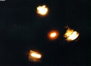
A witness filmed a strange flying object in the sky over Florida
A witness filmed a strange flying object in the sky over Florida
Video of a UFO was taken on the beach.
The author of this video Eric Kipker does not claim with absolute certainty that he was able to remove the alien ship, however, over the Gulf of Mexico that night stuck do something fantastic and inexplicable (see video), which does not like earthly aircraft Esoreiter.
And it is a miracle in that hour was observed by hundreds of eyewitnesses of the resort town of Panama city beach, Florida, no wonder that stuff about this soon became available leading UFO organizations in the United States. Here he writes about it himself Eric Kipker:
“At that moment I was just admiring the sunset over the sea seemed a fantastic picture. And suddenly this picture someone added a little bit of science fiction, UFO, reminiscent of the appearance and behavior of the alien ship. Naturally I was shocked by this event, but managed to remove on video it is a unique heavenly view.”
Ufologists have confirmed the authenticity of the footage however they are confused, what is this apparatus hangs over the Gulf of Mexico, it is possible that he may be just a war toy, a drone or anything else – it is the earthly. But UFO is very similar to the apparatus of the aliens.
So to exclude it from the list of extraterrestrial objects is not, need more data that experts in this field collect. Waiting for new posts about it…
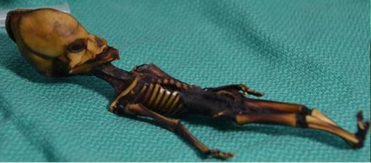
(NEWSER) – A tiny skeleton long rumored to be some kind of alien-human hybrid is actually the body of a stillborn baby girl, researchers say—and the finding has caused outrage in Chile, where the body was dug up near an abandoned Catholic church in 2003. Chilean scientists say it appears that the body was illegally exhumed and smuggled out of the country, making the resulting research deeply unethical, the New York Times reports. The body of the girl, who suffered from severe genetic mutations and bone deformities, ended up in the private collection of Barcelona entrepreneur Ramon Navia-Osorio, who heads an organization for UFO research. Samples were provided to American researchers who reconstructed the genome of the girl, who'd been nicknamed Ata.
Chilean science groups have urged the Genome Research journal to send a message on ethics by retracting the Ata study on the grounds that it's based on the results of grave robbing. "Could you imagine the same study carried out using the corpse of someone's miscarried baby in Europe or America?" the Chilean Society of Biological Anthropology said in a statement. The American researchers are defending the study, saying it has countered claims the skeleton was that of an alien, though they agree the body should be treated with respect. "We now know that it's a child," Stanford University professor of microbiology and immunology Garry Nolan tells the San Jose Mercury News. "I think it should be returned to the country of origin and buried according to the customs of the local people."
Two airline pilots traveling over Arizona claim they were passed by an unidentified flying objects while flying at over 30,000 feet in the air. Both planes reported the sighting and now the Federal Aviation Administration (FAA) has released a radio broadcast of the out-of-this-world incident.
For a seemingly simple fact, the number of organs in the human body is surprisingly controversial. Though the human body hasn’t changed much since modern humans emerged, our total number of organs — or what we call organs — has. A couple of years ago, the number was 78. At the end of 2016, doctors nudged the number up to 79. On Tuesday, scientists announced they’d found the 80th: a network of fluid-transporting vessels that span the entire body called the interstitium. But it has left some people familiar with human anatomy scratching their heads. The interstitium is nothing new.
In a paper published Tuesday in the journal Scientific Reports, doctors propose that the interstitium is the biggest organ in the human body by volume. It’s described as a fluid-filled space, supported by a lattice of collagen fibers, that runs throughout the human body, both within other organs and between them. They don’t call the interstitium an “organ” in the paper, but they’ve referred to it as an organ in interviews with the press, causing some confusion.
This figure from the study shows the interstitium, fluid filled spaces supported by collagen fiber bundles.
At first glance, it seems like they’re just talking about connective tissue, which has long been characterized as the broad class of tissues that support other tissues and organs (some specialized connective tissues are bones, blood, and fat).
What’s new in this study is that the authors are arguing that a specific type of connective tissue is connected within a greater network — the interstitium. This organ, the authors say, may form a protective layer around other organs. Which, admittedly, is huge, if true.
Here’s the summary they provide in their abstract: “In sum, we describe the anatomy and histology of a previously unrecognized, though widespread, macroscopic, fluid-filled space within and between tissues, a novel expansion and specification of the concept of the human interstitium.”
Collagen fibers support the fluid-filled channels that make up the interstitium.
The obvious question is why it took doctors so long to identify the interstitium in the way they just did, and the answer is simple: They just weren’t looking at it right. Previous researchers mischaracterized the connective tissue, the study’s authors say, because once it’s removed from the body for examination, all the fluid flows out, leaving something that looks merely like solid layers of membranes rather than the fluid-filled pockets and channels befitting an organ.
Though some doctors had recently noticed the reticular (that is, net-like) pattern of the interstitium, no one had successfully found a way to sample it. To get past this obstacle, the researchers devised a way to examine bile duct tissue samples obtained during surgeries without losing the structure of the fluid-filled channels. Removing the tissue, they rapidly froze it to retain the reticular structure of collagen fibers and fluid-filled chambers, which are described in the paper.
Their examination revealed that the tissues “are compressible and distensible and may thus serve as shock absorbers.” Furthermore, the channels connect to the lymphatic system, a central part of the human immune system, suggesting it could play a role in immune function. Since it’s a fluid-filled network, it may even function as a highway for cancer cells to travel through the body — a useful map for doctors wanting to understand how cancers metastasize and how to prevent it.
At the moment, though, the implications of this study are far from settled. The truth is that it will require a lot more research to understand what the role of the organ — if it is an organ at all — in the human body is. As The Daily Beastreported, the researchers had the paper rejected more than half a dozen times before Scientific Reports accepted it, so it’s clear that this discovery is a controversial one in the scientific community. That being said, the journal it found a home in is not a fringey rag. As with any medical discovery, time will tell us whether it actually is a discovery.
Abstract:Confocal laser endomicroscopy (pCLE) provides real-time histologic imaging of human tissues at a depth of 60–70 μm during endoscopy. pCLE of the extrahepatic bile duct after fluorescein injection demonstrated a reticular pattern within fluorescein-filled sinuses that had no known anatomical correlate. Freezing biopsy tissue before fixation preserved the anatomy of this structure, demonstrating that it is part of the submucosa and a previously unappreciated fluid-filled interstitial space, draining to lymph nodes and supported by a complex network of thick collagen bundles. These bundles are intermittently lined on one side by fibroblast-like cells that stain with endothelial markers and vimentin, although there is a highly unusual and extensive unlined interface between the matrix proteins of the bundles and the surrounding fluid. We observed similar structures in numerous tissues are subject to intermittent or rhythmic compression, including the submucosae of the entire gastrointestinal tract and urinary bladder, the dermis, the peri-bronchial and peri-arterial soft tissues, and fascia. These anatomic structures may be important in cancer metastasis, edema, fibrosis, and mechanical functioning of many or all tissues and organs. In sum, we describe the anatomy and histology of a previously unrecognized, though widespread, macroscopic, fluid-filled space within and between tissues, a novel expansion and specification of the concept of the human interstitium.
VREEMDE RODE LUCHTEN ZOALS DEZE IN DE OUDHEID VOORKWAMEN...
VREEMDE RODE LUCHTEN ZOALS DEZE IN DE OUDHEID VOORKWAMEN...
Vaak vinden we antwoorden voor het heden en onze nabije toekomst in het verleden, want alles is een cyclus en bepaalde processen blijven zich herhalen.
Door de gloed van de Verwoester was de aarde gehuld in een roodachtig licht. Het land was platgeslagen en verwoest door enorme hagelstenen die neer beukten op alles wat stond.
Het bovenstaande is een klein stukje uit de Kolbrin bijbel. In een eerder artikel schreven wij al over de herkomst van dit werk:
Als we dan uiteindelijk terechtkomen bij iets wat op geschreven documenten lijkt, dan zien we naast de kleitabletten van de Sumeriërs iets wat de Kolbrin Bijbel wordt genoemd. Het is een verzameling van oeroude teksten die vertaald zijn. De herkomst is het oude Egypte en daarna komen we ze weer tegen in de Middeleeuwen.
De manuscripten zouden gered zijn tijdens een brand in het klooster van Glastonbury Abbey in 1184. Er gaat zelfs het gerucht dat de beroemde Nicola Tesla enkele van zijn wijsheden haalde uit de Kolbrin Bijbel. Deze bestaat uit in totaal elf boeken, waarvan men zegt dat de eerste zes geschreven zijn door Egyptenaren ten tijde van de Exodus. Het boek is nooit toegankelijk geweest voor het grote publiek, maar werd altijd geheimgehouden bestemd voor een select groepje mensen. Tegenwoordig is er een gedrukte versie beschikbaar via het Culdian Trust.
Deze Kolbrin bijbel beschrijft in detail de komst van de Verwoester, oftewel de planeet Nibiru of Planet X. In die bijbel staat onder andere het volgende:
De plaag waarde door het land, de rivier was bloedrood en overal was bloed. Zij die dronken van de rivier moesten onmiddellijk overgeven. Het stof beet zich vast in de huid van zowel mens als dier.
Door de gloed van de Verwoester was de aarde gehuld in een roodachtig licht. Het land was platgeslagen en verwoest door enorme hagelstenen die neer beukten op alles wat stond. Ze kwamen naar beneden in hete buien en achter hen aan kwam een soort vreemd gloeiend vuur dat over de aarde liep.
Naarmate Nibiru verder door de ruimte reist, sleurt het als het ware een enorme wolk van ruimtepuin achter zich aan bestaande uit gassen, stenen, rotsen en manen, evenals een enorme wolk van geladen rode roestig stof. Wanneer je daar vanaf de aarde naar kijkt, dan lijkt dit bloedrood. Tijdens de passage van de planeet komt deze wolk dichter in de buurt van de aarde en raakt de enorme staart onze planeet als het ware, waardoor dit roestige stof op aarde terecht komt en de luchten en het water een bloedrode kleur geeft.
Het is ook deze wolk die wij nu regelmatig waarnemen in de vorm van bloedrode zonsondergangen en rode luchten (zelfs 's nachts). Deze rode stofwolk wordt ook door de wind verspreid en zorgt onder ander ook voor bloedrode zandstormen en doet het water op bepaalde plekken eveneens bloedrood kleuren.
Het is niet alleen de Kolbrin bijbel waar deze rode stofwolk wordt beschreven, het komt onder andere ook voor bij de onderzoeker, auteur en goede vriend van Einstein, Immanuel Velikovsky. In zijn boek uit 1950, Werelden in botsing, staat onder andere het volgende:
"Eén van de eerst zichtbare tekenen van deze ontmoeting was het roodkleuren van het aardoppervlak, veroorzaakt door dun stof van roestig pigment. In de zeeën, in de meren en de rivieren zorgde dit pigment voor een bloedrode kleur van het water (pagina 64).
Onze planeet ging dieper in de staart van de komeet. Het stof was de voorbode van het grind (pagina 67).
Net zoals de bekende astronoom Carlos Muñoz Ferrada ging ook Velikovsky ervan uit te maken te hebben met een niet normale planeet, maar een soort komeet/planeet (cometa planeta).
Met al het bovenstaande in het achterhoofd kijken we even naar een aantal gebeurtenissen afgelopen week.
Afgelopen weekend kregen wintersporters in Rusland, Roemenië, Bulgarije en nog een aantal landen te maken met oranje sneeuw. Dit wordt dan verklaart door meteorologen als oranje zand afkomstig van zandstormen in de Sahara.
Maar, deze stormen doen zich niet voor in de ruimte en velen vragen zich dan ook af hoe het kan, als het Sahara zand zou zijn, dat de lucht bij het ruimtestation ISS bloedrood gekleurd is.
Niet alleen rode luchten worden in verband gebracht met de komst van de planeet Nibiru, maar ook paarse zoals wij in een eerder artikel schreven.
Enkele dagen later is de rode gloed bij het ruimtestation veranderd naar een paarse gloed, zoals te zien in de volgende video: Zandstormen komen op die hoogte niet voor. Is er een andere logische verklaring of zien we in de ruimte bij het ISS concrete tekenen van de nadering van Nibiru?
A B-21 Raider Combined Test Force patch has been put up for sale on Ebay. If the insignia is authentic it would be the first indication that such an outfit has been established to support the new stealth bomber's development. It would also be yet another indication that the stealth bomber's development is proceeding swiftly and it fits with recent comments made the of flight test operations at Edwards Air Force Base who noted that his staff "will be testing it here in the near future."
Its basic design consists of yellow ring with black borders and the phrases "B-21" and "Combined Test Force" around a central design. In the middle there is a cloaked figure, which appears to either be the Grim Reaper or another sort of wraith, with a star or "twinkle" in its eye, pointing what may be a skeletal finger that leads, through a trail of smoke, to a silhouette of the B-21.
EBAY/DEANNAANDGORDON
In the background is a globe motif oriented to primarily show the western hemisphere. In red at the top of the globe is the Latin word "praenuntius," which translates to either herald or harbinger, or signs of things to come. Near the bottom there is what may be a bird or dragon in black above the Roman numerals XVII, or 17.
Grim Reapers or similar spectral figures are not uncommon motifs in morale patches, especially those associated with secret programs, those dealing with low-observable designs, or both. "Wraith" is even an informal nickname for the RQ-170 Sentinel.
The star in the eye could be a callback of some sort to the B-2 Combined Test Force logo, which featured a similar heraldic item at the end of one of a pair of bird-of-prey-like talons. If it is indeed a black bird, that could also be related to the names and motifs applied to previous secret and stealth projects, as well.
MILITARYPATCHEANDPINS.COM/THELIVINGMOON.COM
Similar imagery has been used in secretive stealth programs in the past, as well as for the B-2's own CTF.
It's less clear what the Roman numerals "17" might represent. Some have already posited it could be a reference to the actual Combined Test Force squadron, which will be assigned to the 412th Test Wing at Edwards Air Force Base in the future – if it doesn't exist already in some form.
The Wing's 417th Flight Test Squadron shut down operations in 2012 with the cancellation of the YAL-1A airborne laser program and could be the unit in question, but there is no indication the Air Force has reactivated it since then. The Air Force did, without any other apparent notice, reactivate the 452nd Flight Test Squadron at Edwards Air Force Base in March 2017, which seems like a more plausible reference to "17."
It could also have some entirely different meaning altogether. There is an Italian superstition that the Roman numerals for 17 are unlucky since they can form an anagram that translates to "I lived," with the implication that one's life is over.
The patch also caught the eye of our friend David Cenciotti of Theaviaitonist.com, and he says the seller obtained the patch from personnel at Edwards AFB who were standing-up the B-21 test program. This vibes with what The War Zone has long reported—the South Base installation at Edwards AFB has been in transition for some time and that the B-21, and possibly other cutting-edge platforms and munitions that will work with it, will use the facility as a headquarters for development.
We also have to remember that Combined Test Forces work to bring all the elements of a program—including those of contractors and various military stakeholders—together to devise and execute test program that will usher the weapon system into an operational state and then sustain and upgrade it after that. This includes developing and evaluating the many different technologies that make up the aircraft as a whole.
USAF
B-21 Raider
With this in mind, a flying prototype isn't needed for test work to begin, far from it in fact, albeit proof of concept, technology demonstrators, and aircraft that the B-21 borrows tech direct from do likely exist in the classified realm. Still, there are plenty of components to put through their paces before they fly on a finalized design.
Systems integration is one of the biggest challenges during the genesis of any weapons platform, yet alone for one that is likely to end up being the most complex airplane the world has ever seen. We have even heard about some of the issues the bomber has already faced during development.
In other words, the B-21 test team should really exist by now as they have a lot of work to do in a fairly short period of time. The USAF wants the aircraft operationalby around 2025.
The patch has leaped up dramatically in price in recent hours, with a current bid of $103.50. Let us know what you think of its symbolism in the comments below.
Zoomed in and slow mo version of the ufo around the airliner march 2018
Zoomed in and slow mo version of the ufo around the airliner march 2018
A nice, clean, slowed down shot of the ufo captured outside the airliner over turkey that just happened recently, what actually is this, this video gives you a clearer look at the incident
Beste bezoeker, Heb je zelf al ooit een vreemde waarneming gedaan, laat dit dan even weten via email aan Frederick Delaere opwww.ufomeldpunt.be. Deze onderzoekers behandelen jouw melding in volledige anonimiteit en met alle respect voor jouw privacy. Ze zijn kritisch, objectief maar open minded aangelegd en zullen jou steeds een verklaring geven voor jouw waarneming! DUS AARZEL NIET, ALS JE EEN ANTWOORD OP JOUW VRAGEN WENST, CONTACTEER FREDERICK. BIJ VOORBAAT DANK...
Druk op onderstaande knop om je bestand , jouw artikel naar mij te verzenden. INDIEN HET DE MOEITE WAARD IS, PLAATS IK HET OP DE BLOG ONDER DIVERSEN MET JOUW NAAM...
Druk op onderstaande knop om een berichtje achter te laten in mijn gastenboek
Alvast bedankt voor al jouw bezoekjes en jouw reacties. Nog een prettige dag verder!!!
Over mijzelf
Ik ben Pieter, en gebruik soms ook wel de schuilnaam Peter2011.
Ik ben een man en woon in Linter (België) en mijn beroep is Ik ben op rust..
Ik ben geboren op 18/10/1950 en ben nu dus 75 jaar jong.
Mijn hobby's zijn: Ufologie en andere esoterische onderwerpen.
Op deze blog vind je onder artikels, werk van mezelf. Mijn dank gaat ook naar André, Ingrid, Oliver, Paul, Vincent, Georges Filer en MUFON voor de bijdragen voor de verschillende categorieën...
Veel leesplezier en geef je mening over deze blog.