The purpose of this blog is the creation of an open, international, independent and free forum, where every UFO-researcher can publish the results of his/her research. The languagues, used for this blog, are Dutch, English and French.You can find the articles of a collegue by selecting his category. Each author stays resposable for the continue of his articles. As blogmaster I have the right to refuse an addition or an article, when it attacks other collegues or UFO-groupes.
Druk op onderstaande knop om te reageren in mijn forum
Zoeken in blog
Deze blog is opgedragen aan mijn overleden echtgenote Lucienne.
In 2012 verloor ze haar moedige strijd tegen kanker!
In 2011 startte ik deze blog, omdat ik niet mocht stoppen met mijn UFO-onderzoek.
BEDANKT!!!
Een interessant adres?
UFO'S of UAP'S, ASTRONOMIE, RUIMTEVAART, ARCHEOLOGIE, OUDHEIDKUNDE, SF-SNUFJES EN ANDERE ESOTERISCHE WETENSCHAPPEN - DE ALLERLAATSTE NIEUWTJES
UFO's of UAP'S in België en de rest van de wereld Ontdek de Fascinerende Wereld van UFO's en UAP's: Jouw Bron voor Onthullende Informatie!
Ben jij ook gefascineerd door het onbekende? Wil je meer weten over UFO's en UAP's, niet alleen in België, maar over de hele wereld? Dan ben je op de juiste plek!
België: Het Kloppend Hart van UFO-onderzoek
In België is BUFON (Belgisch UFO-Netwerk) dé autoriteit op het gebied van UFO-onderzoek. Voor betrouwbare en objectieve informatie over deze intrigerende fenomenen, bezoek je zeker onze Facebook-pagina en deze blog. Maar dat is nog niet alles! Ontdek ook het Belgisch UFO-meldpunt en Caelestia, twee organisaties die diepgaand onderzoek verrichten, al zijn ze soms kritisch of sceptisch.
Nederland: Een Schat aan Informatie
Voor onze Nederlandse buren is er de schitterende website www.ufowijzer.nl, beheerd door Paul Harmans. Deze site biedt een schat aan informatie en artikelen die je niet wilt missen!
Internationaal: MUFON - De Wereldwijde Autoriteit
Neem ook een kijkje bij MUFON (Mutual UFO Network Inc.), een gerenommeerde Amerikaanse UFO-vereniging met afdelingen in de VS en wereldwijd. MUFON is toegewijd aan de wetenschappelijke en analytische studie van het UFO-fenomeen, en hun maandelijkse tijdschrift, The MUFON UFO-Journal, is een must-read voor elke UFO-enthousiasteling. Bezoek hun website op www.mufon.com voor meer informatie.
Samenwerking en Toekomstvisie
Sinds 1 februari 2020 is Pieter niet alleen ex-president van BUFON, maar ook de voormalige nationale directeur van MUFON in Vlaanderen en Nederland. Dit creëert een sterke samenwerking met de Franse MUFON Reseau MUFON/EUROP, wat ons in staat stelt om nog meer waardevolle inzichten te delen.
Let op: Nepprofielen en Nieuwe Groeperingen
Pas op voor een nieuwe groepering die zich ook BUFON noemt, maar geen enkele connectie heeft met onze gevestigde organisatie. Hoewel zij de naam geregistreerd hebben, kunnen ze het rijke verleden en de expertise van onze groep niet evenaren. We wensen hen veel succes, maar we blijven de autoriteit in UFO-onderzoek!
Blijf Op De Hoogte!
Wil jij de laatste nieuwtjes over UFO's, ruimtevaart, archeologie, en meer? Volg ons dan en duik samen met ons in de fascinerende wereld van het onbekende! Sluit je aan bij de gemeenschap van nieuwsgierige geesten die net als jij verlangen naar antwoorden en avonturen in de sterren!
Heb je vragen of wil je meer weten? Aarzel dan niet om contact met ons op te nemen! Samen ontrafelen we het mysterie van de lucht en daarbuiten.
18-04-2019
Part-Revived Pig Brains Raise Slew of Ethical Quandarie
Part-Revived Pig Brains Raise Slew of Ethical Quandaries
Researchers need guidance on animal use and the many issues opened up by a new study on whole-brain restoration, argue Nita A. Farahany, Henry T. Greely and Charles M. Giattino
Scientists have restored and preserved some cellular activities and structures in the brains of pigs that had been decapitated for food production four hours before. The researchers saw circulation in major arteries and small blood vessels, metabolism and responsiveness to drugs at the cellular level and even spontaneous synaptic activity in neurons, among other things. The team formulated a unique solution and circulated it through the isolated brains using a network of pumps and filters called BrainEx. The solution was cell-free, did not coagulate and contained a haemoglobin-based oxygen carrier and a wide range of pharmacological agents.
The remarkable study, published in this week’s Nature, offers the promise of an animal or even human whole-brain model in which many cellular functions are intact. At present, cells from animal and human brains can be sustained in culture for weeks, but only so much can be gleaned from isolated cells. Tissue slices can provide snapshots of local structural organization, yet they are woefully inadequate for questions about function and global connectivity, because much of the 3D structure is lost during tissue preparation.
The work also raises a host of ethical issues. There was no evidence of any global electrical activity—the kind of higher-order brain functioning associated with consciousness. Nor was there any sign of the capacity to perceive the environment and experience sensations. Even so, because of the possibilities it opens up, the BrainEx study highlights potential limitations in the current regulations for animals used in research.
Most fundamentally, in our view, it throws into question long-standing assumptions about what makes an animal—or a human—alive.
SIGNS OF WHAT?
The pig brains used in the study, which was conducted by a team based largely at Yale School of Medicine in New Haven, Connecticut, produced a flat line on an electroencephalogram (EEG) of brain activity. Had any degree of sentience been recovered, let alone consciousness, one would expect to see low-amplitude waves in the alpha (8–12 Hz) and beta (13–30 Hz) range, at the very least. In consultations with the Neuroethics Working Group of the US National Institutes of Health (NIH) BRAIN Initiative and in discussions with us, the researchers have stated that if they had detected such activity, they would have administered anaesthetic agents to prevent any experience similar to pain or distress, and would have reduced the brain temperature to swiftly quell the activity.
Absence of organized electrical electrical brain activity, measured with an EEG, is one measure used to establish brain death. Credit: Kateryna Kon Getty Images
Researchers already study whole organs, and maintain cellular activity for a few seconds to minutes in slices of animal and human brains. Thus, on the face of it, in the absence of EEG activity, the BrainEx study does not raise fundamentally different issues from those encountered in the use of animal or human brain tissue after death.
Yet, until now, neuroscientists and others have assumed two things. First, that neural activity and consciousness are irretrievably lost within seconds to minutes of interrupting blood flow in mammalian brains. Second, that, unless circulation is quickly restored, there is a largely irreversible progression towards cell death and the death of the organism.
The BrainEx study used pig brains that had received no oxygen, glucose or other nutrients for four hours. As such, it opens up possibilities that were previously unthinkable.
Take the lack of EEG activity. This activity could have been lost irreversibly when the pigs were slaughtered. Another possibility, however, is that the lack of EEG activity was a function of the study design. The researchers used several chemical agents in their solution that inhibit neural activity, hypothesizing that the tissues would be more likely to show some recovery if cellular activity were reduced. Had these blockers been removed at some point, perhaps the team would have detected EEG activity.
Another possibility needing investigation is that something similar to shock treatment for the heart is required to reset the firing of neurons in the brain to a level that is detectable. Or maybe it takes longer than six hours (the length of the BrainEx perfusion, following the four hours after death) for the cells to recover sufficiently for this kind of brain activity to emerge. Physicians sometimes lower the core body temperatures of people who have had a heart attack, to induce a hypothermic coma. This can limit damage caused by swelling in the brain, for instance, and aid cellular recovery. In these cases, patients seem to need at least 24 hours of ‘cooling treatment’.
Obviously, more data are needed, including the replication of the BrainEx findings in other laboratories by other groups. But we’re reminded of a line from the 1987 film The Princess Bride: “There’s a big difference between mostly dead and all dead. Mostly dead is slightly alive.” Even with all the unknowns, the discovery that mammalian brains can be made to seem ‘slightly alive’, hours after the animals had been killed, has implications that ethicists, regulators and society more broadly must now think through.
ANIMAL RESEARCH
To be clear, the BrainEx study did not breach any ethical guidelines for research. The team sought guidance from Yale University’s Institutional Animal Care and Use Committee (IACUC), which exists to ensure that the use of animals aligns with what is required by US law for federally funded research. The committee decided that oversight was unnecessary. The pigs, having been raised as livestock, were exempt from animal welfare laws and were killed before the study started. In the United States, the 1966 Animal Welfare Act is the only federal law that regulates how animals are treated in research, and applies to either living or dead animals. It explicitly excludes animals raised for food. Meanwhile, the policies and regulations of the US Public Health Service, which funds most US research involving animals—mainly through the NIH—do not specify any protections for animals after their death.
Had the research been conducted outside the United States, the response from ethics or regulatory bodies would almost certainly have been the same. The European Union’s Directive on the Protection of Animals Used for Scientific Purposes largely aims to prevent (or minimize) any pain, suffering or distress experienced by live animals. It, too, specifically excludes animals raised for agriculture (see go.nature.com/2cpdgjr). In China, both the Ministry of Science and Technology and the provincial bureaus of science and technology ensure that researchers follow local regulations and that they abide by the National Standard on Laboratory Animal Welfare in China. Here, too, the protections exclude animals raised for food, and the main focus is on eliminating or reducing live animals’ potential pain and distress.
In our view, new guidelines are needed for studies involving the preservation or restoration of whole brains, because animals used for such research could end up in a grey area—not alive, but not completely dead. Five issues in particular need addressing.
First, how should researchers try to detect signs of consciousness or sentience? On its own, EEG activity would not reliably signal a conscious brain; such activity is nearly always detected in people who are under general anaesthesia. EEG activity might provide an appropriate measure should it be detected along with responsiveness to transcranial magnetic stimulation (TMS)—a non-invasive way of stimulating brain activity, using a magnetic coil held near the head. Together with other measures, this would determine the brain’s perturbational complexity index, a way of identifying the level of consciousness. Furthermore, recent research in humans using functional magnetic resonance imaging indicates that certain patterns of neuronal activity may provide a correlate for consciousness.
Second, which species make appropriate models for this type of research on brain perfusion? And what kinds of research and results would be needed to justify the use of other models? (In our view, investigators should proceed cautiously with testing in other mammals, particularly in pigs, dogs or primates, at this time.)
Third, until more is known, is the use of neuronal activity blockers sufficient to safeguard against the emergence of capabilities associated with sentience, such as the capacity to feel pain? It might be necessary to apply BrainEx or similar systems to mice or rats, both with and without neuronal activity blockers, to better understand the blockers’ role.
Fourth, under which scenarios should anaesthetics be used in follow-on studies, to safeguard against the possibility of inducing any experience similar to pain or distress? And under what scenarios might it be permissible not to use them? (We think that the use of anaesthetics in follow-on studies should be mandatory at this time, given all of the unknowns.)
Finally, for how long should BrainEx or similar artificial circulatory systems be run? Such systems might be effective for only a certain period of time, or there could be a limit as to how much recovery can be achieved. This knowledge will inform analyses of risks and benefits.
HUMAN RESEARCH
Although it is a long way off, researchers might one day consider using a system similar to BrainEx to treat humans for brain damage caused by a lack of oxygen. Until now, neuroscientists and physicians have assumed that the cell death caused by this is irreversible. Treatment generally involves working with a person’s remaining healthy brain tissue to help rehabilitate mobility, motor and other skills.
Before developing whole human-brain models outside the body—and certainly before the use of brain perfusion in the clinic—investigators need to arm people with enough information for them to make informed decisions. Most fundamentally, patients or donors will need to understand what kinds of brain activity could result and what that activity could mean. They will also need to know the chances of recovery being only partial, and the implications that will have.
Another question is what information, if any, could plausibly be retrieved from the brain. Various groups are developing ways to decode the neural activity of living people, for instance to probe their memories or the images they have seen in their dreams. Could such approaches one day be applied to brains after death?
Such possibilities (if they come to pass at all) are far in the future. Yet we need to think through at least some of them now. Hundreds of people worldwide have already paid to have their brains frozen and stored, in the hope that scientists will one day be able to revive them. It’s easy to imagine misapplications of brain perfusion following the publication of the BrainEx study alone.
GUIDELINES
It might not be easy for others to replicate the study, despite the BrainEx team providing detailed information on the device, perfusate and methods. As a first step, the investigators, their home institutions and the NIH should facilitate the transfer of the technology and know-how to other researchers and institutions. Any follow-up and independent studies should be just as transparent as this one.
Crucially, future researchers will need guidance through the potential scientific, ethical and political questions opened up by this research.
Precedents exist. Internationally, research involving stem cells derived from human embryos has successfully been steered by the 2005 Guidelines for Human Embryonic Stem Cell Research released by the US Institute of Medicine and US National Research Council—the substance of which was almost entirely adopted by the International Society for Stem Cell Research. Ongoing efforts to set guidelines for human genome-editing research hold lessons, too. Key actors here are the US National Academy of Sciences, the US National Academy of Medicine, the UK Royal Society, the Hong Kong Academy of Sciences, the Chinese Academy of Sciences and the Nuffield Council on Bioethics.
In other contexts, such as in biomedical engineering (see, for example, go.nature.com/2t6kon5), artificial intelligence and debates around the definition of death, international conferences are being held to help find common ground across countries and to develop frameworks that enable responsible scientific progress.
We think that the latest research on brain resuscitation demands the same kind of international attention. A starting point could be the guiding principles issued last December by the Neuroethics Working Group of the NIH BRAIN Initiative, which held a 2018 workshop on research with human neural tissue.
Citizens must be part of the process. Engaging non-scientists in delineating the ethical boundaries of this research doesn’t guarantee its public acceptance in the future; and nor should it, necessarily. But not engaging other stakeholders could help to precipitate its rejection.
In our view, discussion about the appropriate path for this research should not wait for follow-up studies. The Yale group was conscientious and consulted the local institutional IACUC, Yale bioethicists, NIH programme officers and even the NIH Neuroethics Working Group. The researchers did what they could, and probably more than many would have done, to ensure that they were acting appropriately in a void of ethical analysis on the issue.
Now is the time to fill that void.
This article is reproduced with permission and was first published on April 17, 2019.
ABOUT THE AUTHOR(S)
Nita A. Farahany
Nita A. Farahany is professor of law and philosophy at Duke University, director of the Duke Initiative for Science & Society, Duke University, Durham, North Carolina, USA.
Henry T. Greely
Henry T. Greely is Director of the Center for Law and the Biosciences and Professor (by courtesy) of Genetics, at the Stanford School of Medicine. He is the Chair of the Steering Committee of the Center for Biomedical Ethics, and Director of the Stanford Program in Neuroscience and Society.
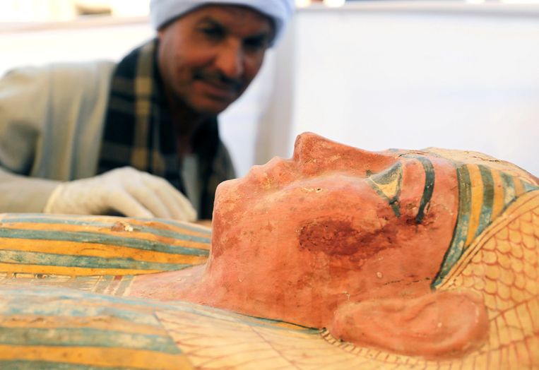
REUTERSEgyptische functionarissen onthulden onder meer een mummie die in Luxor werd ontdekt.
WETENSCHAP Archeologen hebben een 3.500 jaar oud graf blootgelegd en een mummie in een ongeopende kist die van minstens 1.069 voor Christus dateert, op twee sites in de stad Luxor in het zuiden van Egypte. Dit heeft een functionaris voor oudheden vandaag gemeld.
“Het graf van de achttiende dynastie heeft een grote binnenplaats, de grootste die tot dusver in een begraafplaats op deze site werd gevonden”, zei Mostafa Waziri, secretaris-generaal van de Hoge Raad van Oudheden, op een ceremonie op de archeologische site Draa Abul Naga. De necropolis Draa Abul Naga, op de westoever van de Nijl, is bekend om zijn tempels en begraafplaatsen van edellieden uit de oudheid.
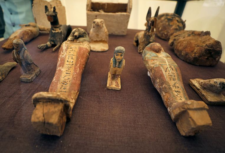
De ploeg van Egyptische archeologen die in augustus 2018 in dit gebied begon te werken, vond ook honderden Oesjabti-beelden, beeldjes die gewoonlijk in grote hoeveelheden in graven worden gevonden.
REUTERSDe beeldjes die door de archeologen werden opgegraven.
Functionarissen onthulden voorts een mummie die elders in Luxor ontdekt werd. De mummie, die in drie ongeopende en in elkaar passende kisten lag, dateert van de Ramessidentijd (1292-1069 voor Christus), die de achttiende en twintigste dynastie omvat.
De onthullingsplechtigheid, op de werelderfgoeddag van Unesco, werd bijgewoond door premier Moustafa Madbouly en minister van Oudheden Khaled al-Anani. Egypte maakte de voorbije maanden een reeks ontdekkingen bekend in de hoop het slabakkende toerisme te doen opleven.
NASAHet ISS, de voeten van Scott Kelly en een uitzicht over de Bahama's.
WETENSCHAPScott Kelly - de astronaut die een jaar in het Internationaal Ruimtestation (ISS) doorbracht om te kijken wat zo’n lang verblijf met het menselijk lichaam doet – leefde zichzelf creatief uit in de ruimte. Met zijn Nikon D4 maakte hij adembenemende foto’s waarvan NASA er nu enkele deelt.
In maart 2015 werd Scott Kelly richting het ISS gestuurd. Daar zou hij een jaar verblijven. De astronaut werd uitgekozen voor de missie omdat hij een eeneiige tweelingbroer heeft. Eenmaal Scott terugkeerde naar de aarde konden wetenschappers uittesten wat voor een invloed de lange ruimtemissie op zijn lijf had. Die invloed bleek overigens behoorlijk groot te zijn: Scott had oogproblemen, geheugenverlies en zijn DNA was niet meer identiek aan dat van zijn broer.
Voordat hij terugkeerde naar de aarde maakte Scott 5.440 toertjes rondom de aarde. Hij zag de zon 10.944 keer opkomen en ondergaan, en hij deelde maar liefst 713 foto’s op Instagram. NASA bundelt er nu een paar, en in het boek van Scott Kelly ‘Infinite Wonder’ worden alle foto’s getoond.
Een ding is alvast duidelijk: de aarde is prachtig (vanuit de ruimte). Zo maakte Scott Kelly een impressionante foto van de kleurrijke Franse kustlijn.
NASADe Franse kustlijn
Ook de Bahama’s lijken te mooi om waar te zijn. “Zo’n verfrissend zicht”, schreef Scott bij de #Bahamas op Instagram.
Ook de foto’s die hij maakte nadat de zon onderging op aarde, zijn net schilderijtjes. New York pik je er meteen uit door de talrijke lichtstraten. Op Twitter deelde Scott trouwens ook een gelijkaardige foto van New York, maar dan eentje waar de zon schijnt. Terwijl de oranje gloed ’s nachts overheerst, ziet de Amerikaanse stad er overdag vooral blauw uit vanuit de ruimte. 7
NASANew York City
Good morning, #NewYorkCity! It’s a great day to be on Earth and in one of my favorite places on the planet! Who’s with me,
0
1
2
3
4
5
- Gemiddelde waardering: 0/5 - (0 Stemmen) Categorie:HLN.be - Het Laatste Nieuws ( NL)
VERNIETIGING GEHEIME BASIS OP DE MAAN ( VIDEO )
VERNIETIGING GEHEIME BASIS OP DE MAAN ( VIDEO )
Wikileaks is de afgelopen week veelvuldig in het nieuws geweest met betrekkingen tot de arrestatie van Julian Assange.
Via diezelfde Wikileaks is er ook onthuld dat de Amerikanen een geheime basis op de maan hebben vernietigd.
Op de website van Scott Waring, de man die dag en nacht speurt op foto’s van NASA, staat een heel interessant verhaalover een geheime basis op de maan.
Het begint met Scott die dit keer in de Wikileaks database aan het snuffelen is en daar een bericht vindt over een geheime basis op de maan die door de Amerikanen zou zijn vernietigd.
Het gaat om het volgende bericht (Klik op het plaatje voor de link naar het originele bericht)
Klaarblijkelijk gaat het hier om een telegram, maar omdat deze handgeschreven is en de inhoud nooit is gedigitaliseerd, kan deze niet online worden bekeken.
En als we denken aan een geheime basis op de maan, dan denken we terug aan de Apollo vlucht die officieel niet bestond en dat is Apollo 20.
Het verhaal is afkomstig van een man die beweert dat hij meegeweest is op een speciale NASA missie: William Rutledge, gepensioneerd en woont nu in Afrika. Hij kwam naarvoren met een aantal verbazingwekkende feiten over zijn betrokkenheid bij NASA aan het eind van de jaren '70.
Rutledge claimt dat hij op tenminste twee missies naar de Maan is geweest, inclusief de mislukte Apollo 19 en de Apollo 20 missie, die naar hij zegt werd gelanceerd in augustus 1976 vanaf de VandenBerg luchtmachtbasis. Deze beide missies waren “geheime gezamenlijke ruimtemissies”, uitgevoerd door de Amerikanen en de Russen. Je vindt ze niet terug op de lijst van officiële missies en als het verhaal waar is, is daar een goede reden voor.
Het doel van deze missies was het onderzoeken van een groot object aan de achterkant van de maan in de Delporte_Iszak regio. Het was voor het eerst ontdekt door de Apollo 15 missie. Het lijkt op een neergestort gigantisch ruimteschip uit oude tijden. Op de volgende foto krijg je ongeveer een idee hoe groot het is:
Scott Waring heeft jarenlang persoonlijk contact gehad met William Rutledge en hij is er dan ook van overtuigd dat de maanbasis waarover in het Wikileaks bericht wordt gesproken, dezelfde is als die waar Rutledge over sprak:
Hier volgen twee van Rutledge afkomstige foto’s van die geheime basis op de maan:
Tijdens hun activiteiten op de maan ontdekten Rutledge en zijn mede astronaut een sigaarvormig ruimteschip in de buurt van de Delporte krater. Zij gingen dit schip binnen door een gat dat zich al aan de zijkant van het ruimtevaartuig bevond en daarbinnen vonden ze glazen buisjes met daarin kleine tweevoetige skeletjes van slechts een paar centimeter lang.
Ook vonden ze daar een vrouwelijke astronaut die later bekend zou worden als de Mona Lisa van de maan. Op het moment dat Rutledge en zijn collega haar vonden was ze in coma en leek mentaal en fysiek verbonden te zijn met het schip.
Ze is nooit meer bij bewustzijn gekomen en ze hebben haar later meegenomen naar de aarde.
Verder ontdekten Rutledge en zijn collega een mooie, maar beschadigde buitenaardse stad op de maan en dit is de stad waarover in het Wikileaks bericht wordt gesproken volgens Scott. Hij denkt dat ze deze basis in een latere missie hebben vernietigd, nadat ze alle mogelijke waardevolle zaken er vandaan hadden gehaald.
In de tijd dat Scott Waring contact had met Rutledge was deze al in de zeventig en vertelde Scott dat hij wat van zijn oude filmmateriaal had omgezet naar digitale video zodat hij dit kon doorgeven voordat hij stierf.
Hieronder volgen enkele van de video’s die Scott heeft gekregen van Rutledge en hij zegt dat ze honderd procent echt zijn.
Nadat Scott het verhaal had gehoord van Rutledge over de Delporte krater is deze wekenlang aan het zoeken geweest en uiteindelijk had hij succes en vond het sigaarvormige schip.
Hoe groot dit schip in werkelijkheid is, wordt duidelijk uit het volgende plaatje.
Toen Scott zijn ontdekking deelde met Rutledge was deze zeer verbaasd over het feit dat NASA deze foto niet had schoon gepoetst en bedankte Scott voor het vertrouwen dat deze altijd in hem had gehad.
Waar Rutledge op dit moment is en of hij nog leeft weet Scott niet, maar wel dat hij ervan overtuigd is dat het bericht van Wikileaks gaat over de stad die Rutledge ontdekte op de maan.
Rocks mysteriously falling from the sky is one of theclassic Fortean phenomenadescribed in Charles Forte’s “Book of the Damned.” Often blamed on poltergeists, pranksters, weather or Bigfoot, these dangerous events are reported frequently in India, which is where the latest occurrence originates. Residents of Raigarh say they now fear nightfall after large rocks have pelted their homes, shops and people since March 2019. With local police not finding any pranksters and India not being a big Bigfoot stomping ground, Raigarheans are suspecting a rock-throwing ghost. Before they call for psychic help, they may want to wait and see if the ghost switches to throwing coins. Wait … what?
“Nobody was found during the search. People in the area live under threat because of this incident. Local people said that it has been happening continuously for five to six days. This makes people in the area feel insecure. The rain of stones starts at seven o’clock in the evening and continues until 11 p.m. Three people were severely injured by stone-pelting on the evening of March 15 due to stones falling on their heads.”
In a Google translation of a report on the media site Patrika.com, residents of Raigarh (a city in east-central India) sound credible and legitimately concerned about the stones and rocks raining down on their village. In the video, they show a pile of the rocks, which they say weigh as much as two kg (4.4 pounds) and appear to all be some kind of quartz. They also show the damage to a house, which is not surprising considering the size of these stones, and report that a number of people have been injured and required medical attention. The local police responsible for the neighborhoods affected have been contacted and bands of young men go out when the rock downpour begins but no human perpetrators have been found.
Ghosts? Stories of poltergeists throwing stones have been told for centuries. A rash of reports in 1682 in New Castle, New Hampshire, gave the phenomena a name: Lithobolia, which is also the name of a 7,000-word narrative folk tale about the event written by Richard Chamberlayne and first printed in 1698 which centers around a stone-throwing devil or demon (the Lithobolia). Stories of spirits throwing stones are a common around the world – there have been recent reports in Zimbabwe, Brazil and another in India. While they could all be blamed on vandals or pranksters, none had concrete proof that the concrete was hurled by humans.
Bigfoot? Those in the Bigfoot research field can call up many examples of the cryptid throwing stones (a common trait of apes as well), and India does have one hairy cryptid – the Mande Burung (jungle man) which is said to resemble a Sasquatch or Yeti and is most often reported in the Meghalaya subtropical forests in the northeastern state of Meghalaya. FYI – that’s about 1,400 km (870 miles) from Raigarh.
It doesn’t appear that Raigarh is in an area prone to seismic activities or rocks falling from mountains. What else could be the cause of its showers of stones? Whatever it is, they may want to wait it out and see if it switches to coins. That’s right … cold, hard cash crashed down from the sky in the Devlochri village of Madhya Pradesh in central India, about 650 km (400 miles) from Raigarh. Patrika.com reports the rain of coins occurred in September 2017 during a thunderstorm. Witnesses reported the coins were of varying denominations and fell in multiple locations. That sounds like a wind or tornado-aided Fortean event like most fish rains turn out to be, but the report had no mentions of a bank or armored car losing a bag of coins to a cyclone. Rich ghosts? Mande Burung celebrating winning the lottery?
Most people in Raigarh would obviously prefer coins over rocks, but what they’d really like are some answers … although 2 kg of rupees would definitely be a nice chunk of change to help pay for the a hole in the roof.
Satellite anomalies and other examples of spaceborne intrigue continue to add up, suggesting that the loomingwar in spacemight be getting closer to hot than we think. In late March 2019, India blew up a satellite in orbit with a missile, littering near-orbits with debris.The display was intended as a show of force for India’s military in the midst of more and more nations testing anti-satellite weaponry and technology.
Shortly after, United States Air Force Secretary Heather Wilson issued a statement which appeared to warn that the U.S. would soon start flexing its muscles in space. At the Space Foundation’s 35th annual Space Symposium this month, Wilson suggested that the Air Force and other armed forces are beginning to shift their focus towards space as the next warfighting domain. With that in mind, Wilson hinted that the U.S. may soon show the world what it’s capable of in space in order to serve as a deterrent for would-be space conflicts:
We looked at all of our missions in space, from missile warning to communications and intelligence collection. We took the best estimates of the threat and presumed a thinking adversary who would respond to the actions that we take. That capability needs to be one that’s understood by your adversary. They need to know there are certain things we can do, at least at some broad level, and the final element of deterrence is uncertainty.
That uncertainty is further hinted at by many recent incidents of strange satellite behavior observed in near-Earth orbit. Earlier this week, satellite tracking firm ExoAnalytic Solutions spotted communications satellite Intelsat 29e behaving erratically. Footage collected with telescopes showed the satellite splintering apart in space as streaks of flammable gasses and debris shot out in all directions. The cause of the satellite’s demise remains unknown, but ExoAnalytic Solutions is currently investigating the incident.
Like all unmanned aircraft, satellites continue to get smaller and smaller, opening up new possibilities for weaponry.
A few days later, astronomers at the Russia-based Institute of Solar-Terrestrial Physics spotted an anomalous satellite in orbit which appears to be maneuvering between other satellites. Russia, China, and the US have in recent years launched experimental and largely secret micro-satellites which are believed to be able to hijack or manipulate adversaries’ satellites and monitor their communications first-hand. It’s unknown who might be controlling this particular satellite, but of course Russian government mouthpiece Sputnik Newsalleges that it’s the Americans as they always do. But hey, maybe it is. It probably is.
While science fiction has for decades predicted that the next war would happen in space, the fact that many of the superpowers are already testing space weapons shows that art indeed often becomes reality. I’m left to wonder how many of the current anomalous phenomena being observed may be a byproduct of this new weapons testing.
Move over, ‘Oumuamua … there’s a new interstellar object in town. Well, there was, according to Abraham (Avi) Loeb, the astronomer who will be forever linked to ‘Oumuamua because of his theory that the cigar-shaped, not-of-this-solar-system object might actually be artificially made – in other words, an interstellar spaceship or solar sail. Despite some ridicule, Harvard didn’t fire the chair of its Astronomy Department and Loeb went back to work … and has now discovered his own interstellar object which predates ‘Oumuamua. Avi, can you give this one a name that’s easier to spell and pronounce?
“I was very surprised. I didn’t expect that. I thought we will not see anything. But in retrospect, like any discovery, you say, Oh yeah, of course. How could I be so foolish not to look for that in the first place?”
“That” is — or was – a meteor that was reported over Manus Island in Papua New Guinea on January 8, 2014. In a paper that has been submitted to The Astrophysical Journal Letters, Loeb and Harvard undergraduate student Amir Siraj describe how they found the report while reviewing the Center for Near-Earth Object Studies’ catalog of meteor events for objects that had two key ingredients indicating they might be from another galaxy – speed and trajectory.
“We know the motion of the Earth [and] we correct for it—for the gravity of Earth, gravity of the sun, gravity of all the planets.”
Loeb told National Geographic that he and Siraj searched through 30 years of data and found one meteor that had the right combination of both. The meteor was traveling at almost 37 miles per second (134,200 mph or 216,000 km/h) when it disintegrated over Manus Island. That’s too fast to have been slingshot at Earth by a tight loop around the Sun or Jupiter or another planet in the way NASA gives space probes a boost in velocity, which means it was probably fired at us by another star. While there are no pictures of this meteor, the data in the CNEOS catalog indicated an unusual trajectory that, when coupled with its speed, indicated this was most likely an interstellar object that arrived three years before ‘Oumuamua.
“If we identified such a thing in real time, we could take a spectrum and figure out the composition.”
Unfortunately, this one burned up in the atmosphere due to its size — three feet across and weighing about 1,100 pounds. And no, it wasn’t cigar-shaped, so Loeb doesn’t think it was a spaceship. However, it could still have been carrying life forms.
“You can imagine that if these meteors were ejected from the habitable zone of a star, they could help transfer life from one planetary system to another.”
Panspermia! Loeb thinks most of these interstellar objects are much smaller than ‘Oumuamua (1 km long) and will disintegrate before impacting, but much could be learned by beefing up the NEO detection system to catch more of them as they burn up in the atmosphere so their spectrums can be analyzed for mineral content.
That’s not as exciting as finding an alien spaceship, but Avi Loeb seems well on his way to becoming the go-to astronomer for interstellar objects.
A Great Ancient Mystery Never Fully Understood Until No is Finally Solved
A Great Ancient Mystery Never Fully Understood Until Now is Finally Solved
This is an amazing immersion in the ascension teachings of the mystical ancients, including the Sumerians, the Egyptians, the Essenes and the Cathars of Southern France.
Transformation into light being was their goal. William presents persuasive evidence that certain mystics knew the secrets of the Book of Love for activating the spiritual capability of our spiritual DNA and connecting with the “light beyond light” that transforms us into luminous beings (angels) on contact.
This book is the highest teaching of the ancient angels and aliens. A prophecy says the Book of Love would be discovered at a preordained time…our time.
Its power would be so great that all anger, jealousy and fear would vanish from our world. In this beautifully illustrated presentation, you’ll discover techniques for manifesting the Book of Love. More, you’ll discover the next best version of yourself.
Real Alien Human Hybrid Programs - A New Race is Appearing on Earth
Real Alien Human Hybrid Programs -A New Race is Appearing on Earth
In this complimentary episode from George Noory’s Gaia TV show, Beyond Belief, hypnotherapist Barbara Lamb details how many of her clients have had encounters with otherworldly beings; and a number of those have had startling similar experiences which revealed they’d been subjected to extraterrestrial hybridization programs.
0
1
2
3
4
5
- Gemiddelde waardering: 0/5 - (0 Stemmen) Categorie:ALIEN LIFE, UFO- CRASHES, ABDUCTIONS, MEN IN BLACK, ed ( FR. , NL; E )
Linda Moulton Howe Something is in the Solar System Sending a Message
Linda Moulton Howe Something is in the Solar System Sending a Message
COAST TO COAST AM. Investigative reporter Linda Moulton Howe delved into radio bursts over a billion light-years from Earth, the decline of coral in Australia’s Great Barrier Reef, and an account of a huge underground pyramid and its link to alien influences.
In January, scientists announced that very rapid radio bursts were mysteriously repeating, which led to the question: Could fast radio bursts be powering alien probes or even interstellar transports with heavy payloads up to a million tons? She spoke with Harvard’s Astronomy Chair, Avi Loeb, who believes such a theory is possible.
The source behind such radio bursts would have to be immensely powerful, which suggests it could be artificial, he indicated, adding that an Earth-sized radiowave beam focused on a huge “light sail” could possibly power interstellar cargo craft.
Scientists Restore Some Functions in a Pig’s Brain Hours after Death
Scientists Restore Some Functions in a Pig’s Brain Hours after Death
Circulation and cellular activity were restored in a pig’s brain four hours after its death, a finding that challenges long-held assumptions about the timing and irreversible nature of the cessation of some brain functions after death, Yale scientists report April 17 in the journal Nature.
The brain of a postmortem pig obtained from a meatpacking plant was isolated and circulated with a specially designed chemical solution. Many basic cellular functions, once thought to cease seconds or minutes after oxygen and blood flow cease, were observed, the scientists report.
Immunofluorescent stains for neurons (green), astrocytes (red), and cell nuclei (blue) in a region of the hippocampus of a pig’s brain left untreated 10 hours after death (left) or subjected to perfusion with the BrainEx technology. Ten hours postmortem, neurons and astrocytes undergo cellular disintegration unless salvaged by the BrainEx system.
Image credit: Stefano G. Daniele & Zvonimir Vrselja; Sestan Laboratory; Yale School of Medicine
“The intact brain of a large mammal retains a previously underappreciated capacity for restoration of circulation and certain molecular and cellular activities multiple hours after circulatory arrest,” said senior author Nenad Sestan, professor of neuroscience, comparative medicine, genetics, and psychiatry.
However, researchers also stressed that the treated brain lacked any recognizable global electrical signals associated with normal brain function.
“At no point did we observe the kind of organized electrical activity associated with perception, awareness, or consciousness,” said co-first author Zvonimir Vrselja, associate research scientist in neuroscience. “Clinically defined, this is not a living brain, but it is a cellularly active brain.”
Cellular death within the brain is usually considered to be a swift and irreversible process. Cut off from oxygen and a blood supply, the brain’s electrical activity and signs of awareness disappear within seconds, while energy stores are depleted within minutes. Current understanding maintains that a cascade of injury and death molecules are then activated leading to widespread, irreversible degeneration.
However, researchers in Sestan’s lab, whose research focuses on brain development and evolution, observed that the small tissue samples they worked with routinely showed signs of cellular viability, even when the tissue was harvested multiple hours postmortem. Intrigued, they obtained the brains of pigs processed for food production to study how widespread this postmortem viability might be in the intact brain. Four hours after the pig’s death, they connected the vasculature of the brain to circulate a uniquely formulated solution they developed to preserve brain tissue, utilizing a system they call BrainEx. They found neural cell integrity was preserved, and certain neuronal, glial, and vascular cell functionality was restored.
The new system can help solve a vexing problem — the inability to apply certain techniques to study the structure and function of the intact large mammalian brain — which hinders rigorous investigations into topics like the roots of brain disorders, as well as neuronal connectivity in both healthy and abnormal conditions.
“Previously, we have only been able to study cells in the large mammalian brain under static or largely two-dimensional conditions utilizing small tissue samples outside of their native environment,” said co-first author Stefano G. Daniele, an M.D./Ph.D. candidate. “For the first time, we are able to investigate the large brain in three dimensions, which increases our ability to study complex cellular interactions and connectivity.”
While the advance has no immediate clinical application, the new research platform may one day be able to help doctors find ways to help salvage brain function in stroke patients, or test the efficacy of novel therapies targeting cellular recovery after injury, the authors say.
The research was primarily funded by the National Institutes of Health’s (NIH) BRAIN Initiative.
“This line of research holds hope for advancing understanding and treatment of brain disorders and could lead to a whole new way of studying the postmortem human brain,” said Andrea Beckel-Mitchener, chief of functional neurogenomics at the NIH’s National Institute of Mental Health, which co-funded the research.
The researchers said that it is unclear whether this approach can be applied to a recently deceased human brain. The chemical solution used lacks many of the components natively found in human blood, such as the immune system and other blood cells, which makes the experimental system significantly different from normal living conditions. However, the researcher stressed any future study involving human tissue or possible revival of global electrical activity in postmortem animal tissue should be done under strict ethical oversight.
“Restoration of consciousness was never a goal of this research,” said co-author Stephen Latham, director of Yale’s Interdisciplinary Center for Bioethics. “The researchers were prepared to intervene with the use of anesthetics and temperature-reduction to stop organized global electrical activity if it were to emerge. Everyone agreed in advance that experiments involving revived global activity couldn’t go forward without clear ethical standards and institutional oversight mechanisms.”
There is an ethical imperative to use tools developed by the Brain Initiative to unravel mysteries of brain injuries and disease, said Christine Grady, chief of the Department of Bioethics at the NIH Clinical Center.
“It’s also our duty to work with researchers to thoughtfully and proactively navigate any potential ethical issues they may encounter as they open new frontiers in brain science,” she said.
Contacts and sources: Bill Hathaway Yale University
Citation: Restoration of brain circulation and cellular functions hours post-mortem. Vrselja, Z. et al. Nature, 2019 DOI: 10.1038/s41586-019-1099-1
0
1
2
3
4
5
- Gemiddelde waardering: 0/5 - (0 Stemmen) Categorie:SF-snufjes }, Robotics and A.I. Artificiel Intelligence ( E, F en NL )
Lost Ancient Civilizations Documentary 2019 Cities Beneath the Jungles, Deserts and Seas
Lost Ancient Civilizations Documentary 2019 Cities Beneath the Jungles, Deserts and Seas
There are many ancient mysteries in human history, none capture the attention as much as long lost civilisations that have never been rediscovered.
In a new, never seen before documentary, we will look in deserts, dense jungles and even underwater and prove that ancient cities are just waiting to be found. With the advance of new technology, it is entirely possible that archaeologists will one day make a history changing discovery that will simply defy comprehension, by unearthing a previously unknown civilisation.
Watch eye-opening documentaries by subscribing and of course hit the bell button in the top right to stay informed of our latest releases. Leave a like, comment and of course share far and wide.
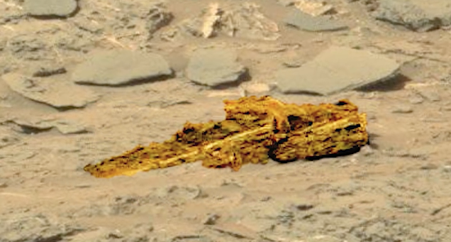
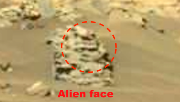
NASA Rover Finds Half-Buried Machine on Mars April 2019, Video, UFO Sighting News.
NASA Rover Finds Half-Buried Machine on Mars April 2019, Video, UFO Sighting News.
Date of discovery: April 17, 2019 Location of discovery: Mars Source photo: unknown In this video Youtube TheRealJimmyRoberts1 shows us alien artifacts that no one knew was even there. He has a great eye for details hidden among the mars rocks and debris. This is just one incredible video that you don't want to miss. Scott Waring
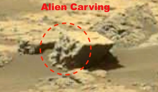
Youtube WhatsUpInTheSky37 found some remarkable alien artifacts on Mars in a NASA photo. He has a great eye for detail and as you see in the photo above, there is an alien figure carved into the stone. The carvings appear to be a bit similar to those of the ancient Aztecs here on Earth. Very exciting discoveries and absolute proof that intelligent aliens once lived on Mars.
How much evidence do you need before you accept the idea that Mars was once teeming with microbial life forms? Would two meteorites sent direct from the surface of the Red Planet that are filled with fossils convince you? Then get ready to yell “I believe!” Scientists given permission to take a thin slice out of a proven Martian meteorite have discovered “mineralized biosignatures” – minerals altered by the presence of microbes and their mineralized fossils. Does this call for champagne or a Martian red wine?
“Comparing recent results and interpretation with other meteorites, it can be raised, that on these similarities the microbially mediated biosignatures can be proposed microbial mediation by FeOB on Mars.”
“FeOB” stands for “iron (FE)-oxiding bacteria and it was discovered in the meteorite ALH-77005 Shergottite discovered in Allan Hills in Antarctica in 1977 and named for the Shergotty meteorite found on the planes of Sherghati, India in 1865 and subsequently proven to be the first known meteorite that was once a part of Mars. In their paper published in Open Astronomy (with pictures), Ildiko Gyollai, Márta Polgári and Szaniszló Bérczi from the Hungarian Academy of Science (HAS) Research Centre for Astronomy and Earth Sciences described how they used optical microscopy and infrared technology along with isotope tests to identify signs of bacteria which survive by eating iron rust – FeOBs.
Hold on! How do they know Earth FeOBs didn’t leave their signatures on this meteorite after it landed? Good question, but the team is way ahead of you.
“The microbial alteration occur only in recrystallized shock melt pocket and near to opaque minerals. Microbial mediation along rims and fractures of coarse grains are not present, which dismiss the terrestrial alteration origin.”
So, the key indicators do not appear near rims and fractures that would allow them in after the meteorite arrived on Earth. Were any of the microbes alive when the chunk left Mars? That’s possible, but the impact that knocked the large piece out of the Red Planet that became one or more meteors occurred four billion years ago, while the meteorites didn’t come to Earth until about 13,000 years ago.
Researchers may want to go back and check old meteor sites for evidence of microbial signatures
Reports on the study point out that this is the second Martian meteorite to show signs of life. The first, ALH-84001, also came from the Antarctic Allan Hills and microbial indicators were discovered in it in 1996. They also note that skeptics say the features could have non-organic origins, but neither can be proven conclusively.
There’s only one way to do that – more Martian mining rovers or hole-digging humans.
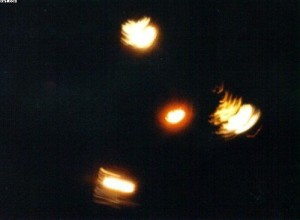
Are aliens hiding a city-sized space station behind our moon, just peeping around the back of it from time to time, in broad daylight and clear skies, to see what we’re up to? That’s what a video posted online recently claims to show. As I’vepostulated before, maybe aliens are trying desperately to get our attention but we’re so thick-headed and resigned to disbelief that they’re forced to keep upping the ante. They tried crop circles, but no one cares about corn. They tried blowing up our cows, but no one cares about cows. They tried shutting off our nukes, but apparently no one cares about nukes either. They’ve sent downmothman, bigfoot, the men in black, time and time again and the people of earth see these impossible things and say “nah.” Alright, they think, time to bring out the big guns. Show ’em the mothership.
The video in question was posted to YouTube by a user named Danil Tumanny—you can watch it here—and gained traction when it was picked up by the popular YouTube channel UFOmania. It seems to show an impossibly huge UFO flying near the moon and disappearing behind it. The UFO looks like a classic doughnut shaped space station like you would see in nearly every piece of science fiction. It’s also what a real giant space station would probably look like. The video was shot in broad daylight, in a cloudless sky. The camera shake is reduced to a minimum, while still being within the limits legally required for a UFO video.
It looks like this, basically.
It sure does look like a big ring of something weird flying around the backside of our beloved satellite. But, like always, there’s a few problems with this video. First, the video is title “123,” has no description, and is tagged in the category “music.” There is no music. We have no details on where, when, or how this video was shot, so there’s no way to check if anyone else saw something strange.
But that shouldn’t really be an issue considering problem number two: If a ship the size of New York City was flying around the moon, in broad daylight, someone else would have seen it. He’s not in the middle of nowhere, you can hear traffic in the background of the video. There’s 7.53 billion people on this planet, and no one else said “well would you look at that?” Maybe the aliens need to up the ante a bit more, because apparently no one cares about city sized spaceships orbiting the moon, either.
Maybe something like this is all that will get our attention.
Lastly, when the ship crosses behind the moon it looks like that edge of the moon sort of flattens out as the ship crosses behind it, which would be proof that it is computer generated. I could be wrong, but this video just doesn’t seem authentic. I don’t want to call it a hoax, because I don’t think that’s what it is. I think a dude was testing out his video editing skills and uploaded the video to YouTube, probably to show a friend or collaborator and never meant for it to gain traction—hence the “123” title and the very wrong category. The video itself doesn’t claim to show anything.
Which makes me a bit more comfortable, honestly. If there was a giant doughnut orbiting the moon and still no one cared, we’d have some big problems on our hands.
They say that truth is stranger than fiction. And they are right – whoever “they” actually are. For me, at least, this really hit home on an expedition I made to Puerto Rico in 2010. It turned out to be one of the most memorable, and strangest, of all my many treks around Puerto Rico, in search of the Chupacabra. It was a Thursday afternoon in January, and, as so often happens, I got a telephone call out of the blue from a television production company that was interested in having me on a new documentary that was just about to be filmed. One of the first questions that the researcher asked me went something like this: “Do you know of any witnesses to the Chupacabra who we can speak to?” Well, yes, I did. Plenty of them. I still do! One of the witness was a woman named Guanina. I met her several years earlier – on a previous TV shoot on Puerto Rico. She lived in Moca, situated on the west side of the island, and which was founded in 1772 by oneDon Jose de Quinonez.
It had been some time since I had first briefly met Guanina, but I told Mark – the guy behind the show – I would give her a call and see if we could get things moving. Fortunately, she remembered me; we had a laugh and a joke on the phone about how, on my earlier trip, all the kids in the neighborhood had come running out to see what was going on when we did a bit of background filming on the outskirts of Moca. Guanina had been a godsend: she and her husband ran a small café and had generously supplied us with plenty of water and soda, as the cameras rolled under a merciless sun. We were soon to interview Guanina. Mark thought we were going to get a definitive tale of the Chupacabra. What he got, however, was something very different – but something very intriguing, too.
Around 2:00 p.m. on the afternoon of our first day of investigation, Mark and I arrived at the café. Guanina gave me a big hug, thrust a cold can of Sprite into my hand, and dished up for me a great meal. Guanina, forty-something, dark-haired and tanned, had an interesting theory about a creature that, back in the mid-1970s, became briefly legendary in the area. It was known as the Moca Vampire, a winged, blood-sucking monster. It was a theory, however, that dated back to around 1987. Mark, barely able to contain himself, eagerly set up his camera and started rolling, as Guanina and I settled back in our chairs to chat. A teenager back in 1987, Guanina used to enjoy walking the hills around Moca. That is, however, until a decidedly traumatic, and even horrific, experience occurred in May 1987 and put paid to all of that. As she strolled around the pathways, Guanina suddenly heard the unmistakable screech of a pig in distress. She raced up the hill, for a further forty or fifty feet or so, and was confronted by a shocking sight: six or seven monkeys were viciously attacking the poor pig, which, by now, was on the ground and clearly close to being in mortal danger.
Guanina shouted at the monkeys, which suddenly ceased their attack, and turned their eyes away from the pig and onto Guanina. For a second or two, there was a tense stand-off. Fortunately, however, the monkeys merely made violent, screaming chatter and then raced away into the deeper grass of the hill. Equally fortunately, the pig – although obviously traumatized, but not physically hurt – unsteadily rose to its feet, stood around for a few minutes, presumably trying to get its bearings, and then wandered off into the undergrowth. Not surprisingly, a terrified Guanina raced down the hill to the safety of her home.
When Guanina told her parents what she had just seen, all three decided to look into the matter further. Scanning various books in the local library, they were soon able to identify the attacking animals as Rhesus monkeys. There were, however, two things that quite rightly puzzled the family: although there are hundreds of Rhesus monkeys on the nearby island of Cayo Santiago – at the Caribbean Primate Research Center – there should not have been any on mainland Puerto Rico. Plus, Rhesus monkeys live chiefly on fruit, cereal and seeds. Occasionally, they will eat bugs and grubs. They are not, however, noted for launching concerted, savage attacks on fully grown pigs. Or, more correctly, normal Rhesus monkeys aren’t known for doing that.
The story was a great one, and Mark was practically ecstatic about the possibility that the Chupacabras were really crazed monkeys. He was also enthused by Guanina’s theory that the 1975 “Moca Vampire” wave was also prompted by attacks by dangerous monkeys. But, the footage never made the cut. Just another few days of memorable, monstrous weirdness!
The late Award-winning journalist and New York Times Best Selling author Jim Marrs presented a program on what the military-trained remote viewers experienced when using this mental technology to study alien life forms at the 2016 Ozark Mountain UFO Conference.
Remote Viewing, a psychic technique developed and utilized by the U.S. Army, may have been instrumental in ending the Cold War as there were no longer any secrets thanks to the psychic experiments on the part of both the Soviets and the U.S.
This practice has since been used to take a look at the different extraterrestrial species interacting with the Earth.
0
1
2
3
4
5
- Gemiddelde waardering: 0/5 - (0 Stemmen) Categorie:ALIEN LIFE, UFO- CRASHES, ABDUCTIONS, MEN IN BLACK, ed ( FR. , NL; E )
New Device Creates Electricity from Falling Snow: It's Small Cheap and Flexible
New Device Creates Electricity from Falling Snow: It's Small Cheap and Flexible
UCLA researchers and colleagues have designed a new device that creates electricity from falling snow. The first of its kind, this device is inexpensive, small, thin and flexible like a sheet of plastic.
Credit: CC0 Public Domain
“The device can work in remote areas because it provides its own power and does not need batteries,” said senior author Richard Kaner, who holds UCLA’s Dr. Myung Ki Hong Endowed Chair in Materials Innovation. “It’s a very clever device — a weather station that can tell you how much snow is falling, the direction the snow is falling, and the direction and speed of the wind.”
The researchers call it a snow-based triboelectric nanogenerator, or snow TENG. A triboelectric nanogenerator, which generates charge through static electricity, produces energy from the exchange of electrons.
Hiking shoe with device attached
Credit Abdelsalam Ahmed
“Static electricity occurs from the interaction of one material that captures electrons and another that gives up electrons,” said Kaner, who is also a distinguished professor of chemistry and biochemistry, and of materials science and engineering, and a member of the California NanoSystems Institute at UCLA. “You separate the charges and create electricity out of essentially nothing.”
Snow is positively charged and gives up electrons. Silicone — a synthetic rubber-like material that is composed of silicon atoms and oxygen atoms, combined with carbon, hydrogen and other elements — is negatively charged. When falling snow contacts the surface of silicone, that produces a charge that the device captures, creating electricity.
“Snow is already charged, so we thought, why not bring another material with the opposite charge and extract the charge to create electricity?” said co-author Maher El-Kady, a UCLA assistant researcher of chemistry and biochemistry.
“While snow likes to give up electrons, the performance of the device depends on the efficiency of the other material at extracting these electrons,” he added. “After testing a large number of materials including aluminum foils and Teflon, we found that silicone produces more charge than any other material.”
About 30 percent of the Earth’s surface is covered by snow each winter, during which time solar panels often fail to operate, El-Kady noted. The accumulation of snow reduces the amount of sunlight that reaches the solar array, limiting the panels’ power output and rendering them less effective. The new device could be integrated into solar panels to provide a continuous power supply when it snows, he said.
The device can be used for monitoring winter sports, such as skiing, to more precisely assess and improve an athlete’s performance when running, walking or jumping, Kaner said. It also has the potential for identifying the main movement patterns used in cross-country skiing, which cannot be detected with a smart watch.
It could usher in a new generation of self-powered wearable devices for tracking athletes and their performances.
Credit: Thom Holmes/Unsplas
It can also send signals, indicating whether a person is moving. It can tell when a person is walking, running, jumping or marching.
The research team used 3-D printing to design the device, which has a layer of silicone and an electrode to capture the charge. The team believes the device could be produced at low cost given “the ease of fabrication and the availability of silicone,” Kaner said. Silicone is widely used in industry, in products such as lubricants, electrical wire insulation and biomedical implants, and it now has the potential for energy harvesting.
Co-authors include Abdelsalam Ahmed, who conducted the research while completing his doctoral studies at the University of Toronto; Islam Hassan and Ravi Selvaganapathy of Canada’s McMaster University; and James Rusling of the University of Connecticut and his research team.
Kaner’s research was funded by Nanotech Energy, a company spun off from his research (Kaner is chair of its scientific advisory board and El-Kady is chief technology officer); and Kaner’s Dr. Myung Ki Hong Endowed Chair in Materials Innovation.
Kaner’s laboratory has produced numerous devices, including a membrane that separates oil from water and cleans up the debris left by oil fracking. Fracking is a technique to extract gas and oil from shale rock.
Kaner is among the world’s most influential and highly cited scientific researchers. He was selected as the recipient of the American Institute of Chemists 2019 Chemical Pioneer Award, which honors chemists and chemical engineers who have made outstanding contributions that advance the science of chemistry or greatly impact the chemical profession.
Contacts and sources:
Stuart Wolpert / University of California – Los Angeles (UCLA)
Citation:
All printable snow-based triboelectric nanogenerator. Abdelsalam Ahmed, Islam Hassan, Islam M. Mosa, Esraa Elsanadidy, Gayatri S. Phadke, Maher F. El-Kady, James F. Rusling, Ponnambalam Ravi Selvaganapathy, Richard B. Kaner. Nano Energy, 2019; 60: 17 DOI: 10.1016/j.nanoen.2019.03.032
0
1
2
3
4
5
- Gemiddelde waardering: 0/5 - (0 Stemmen) Categorie:SF-snufjes }, Robotics and A.I. Artificiel Intelligence ( E, F en NL )
TESS Finds Earth-Sized Planet Orbiting Nearby Star and It's a Hottie
TESS Finds Earth-Sized Planet Orbiting Nearby Star and It's a Hottie
NASA’s Transiting Exoplanet Survey Satellite, TESS, has discovered its first Earth-sized exoplanet. The planet, named HD 21749c, is the smallest world outside our solar system that TESS has identified yet.
In a paper published today in the journal Astrophysical Journal Letters, an MIT-led team of astronomers reports that the new planet orbits the star HD 21749 — a very nearby star, just 52 light years from Earth. The star also hosts a second planet — HD 21749b — a warm “sub-Neptune” with a longer, 36-day orbit, which the team reported previously and now details further in the current paper.
The new Earth-sized planet is likely a rocky though uninhabitable world, as it circles its star in just 7.8 days — a relatively tight orbit that would generate surface temperatures on the planet of up to 800 degrees Fahrenheit.
NASA’s Transiting Exoplanet Survey Satellite (TESS), shown here in a conceptual illustration, will identify exoplanets orbiting the brightest stars just outside our solar system.
Image: NASA’s Goddard Space Flight Center
The discovery of this Earth-sized world is nevertheless exciting, as it demonstrates TESS’ ability to pick out small planets around nearby stars. In the near future, the TESS team expects the probe should reveal even colder planets, with conditions more suitable for hosting life.
“For stars that are very close by and very bright, we expected to find up to a couple dozen Earth-sized planets,” says lead author and TESS member Diana Dragomir, a postdoc in MIT’s Kavli Institute for Astrophysics and Space Research. “And here we are — this would be our first one, and it’s a milestone for TESS. It sets the path for finding smaller planets around even smaller stars, and those planets may potentially be habitable.”
TESS has been hunting for planets beyond our solar system since it launched on April 18, 2018. The satellite is a NASA Astrophysics Explorer mission that is led and operated by MIT, and is designed to observe nearly the entire sky, in overlapping, month-long patches, or “sectors,” as it orbits the Earth. As it circles our own planet, TESS focuses its four cameras outward to monitor the nearest, brightest stars in the sky, looking for any periodic dips in starlight that could indicate the presence of an exoplanet as it passes in front of its host star.
Over its two-year mission, TESS aims to identify for the astronomy community at least 50 small, rocky planets, along with estimates of their masses. To date, the mission has discovered 10 planets smaller than Neptune, four of their masses which have been estimated, including π Men b, a planet twice the size of Earth, with a six-day orbit around its star; LHS 3844b, a hot, rocky world that’s slightly bigger than Earth and circles its star in a blistering 11 hours; and TOI 125b and c — two “sub-Neptunes” that orbit the same star, both within about a week. All four of these planets were identified from data obtained during TESS’ first two observing sectors — a good indication, the team writes in its paper, that “many more are to be found.”
Dragomir picked out this newest, Earth-sized planet from the first four sectors of TESS observations. When these data became available, in the form of light curves, or intensities of starlight, she fed them into a software code to look for interesting, periodic signals. The code first identified a possible transit that the team later confirmed as the warm sub-Neptune they announced earlier this year.
As is usually the case with small planets, where there’s one, there are likely to be more, and Dragomir and her colleagues decided to comb through the same observations again to see if they could spot any other small worlds hiding in the data.
“We know these planets often come in families,” Dragomir says. “So we searched all the data again, and this small signal came up.”
The team identified a small dip in the light from HD 21749, that occurred every 7.8 days. Ultimately, the researchers identified 11 such periodic dips, or transits, and determined that the star’s light was being momentarily blocked by a planet about the size of the Earth.
While this is the first Earth-sized planet discovered by TESS, other Earth-sized exoplanets have been discovered in the past, mainly by NASA’s Kepler Space Telescope, a since-retired telescope that monitored more than 530,000 stars. In the end, the Kepler mission detected 2,662 planets, many of which were Earth-sized, and a handful of those were deemed to be within their star’s habitable zone — where a balance of conditions could be suitable for hosting life.
However, Kepler observed stars that are many leagues further away than those that are monitored by TESS. Therefore, Dragomir says that following up on any of Kepler’s far-flung, Earth-sized planets would be much harder than studying planets orbiting TESS’ much closer, brighter stars.
“Because TESS monitors stars that are much closer and brighter, we can measure the mass of this planet in the very near future, whereas for Kepler’s Earth-sized planets, that was out of the question,” Dragomir says. “So this new TESS discovery could lead to the first mass measurement of an Earth-sized planet. And we’re excited about what that mass could be. Will it be Earth’s mass? Or heavier? We don’t really know.”
Beste bezoeker, Heb je zelf al ooit een vreemde waarneming gedaan, laat dit dan even weten via email aan Frederick Delaere opwww.ufomeldpunt.be. Deze onderzoekers behandelen jouw melding in volledige anonimiteit en met alle respect voor jouw privacy. Ze zijn kritisch, objectief maar open minded aangelegd en zullen jou steeds een verklaring geven voor jouw waarneming! DUS AARZEL NIET, ALS JE EEN ANTWOORD OP JOUW VRAGEN WENST, CONTACTEER FREDERICK. BIJ VOORBAAT DANK...
Druk op onderstaande knop om je bestand , jouw artikel naar mij te verzenden. INDIEN HET DE MOEITE WAARD IS, PLAATS IK HET OP DE BLOG ONDER DIVERSEN MET JOUW NAAM...
Druk op onderstaande knop om een berichtje achter te laten in mijn gastenboek
Alvast bedankt voor al jouw bezoekjes en jouw reacties. Nog een prettige dag verder!!!
Over mijzelf
Ik ben Pieter, en gebruik soms ook wel de schuilnaam Peter2011.
Ik ben een man en woon in Linter (België) en mijn beroep is Ik ben op rust..
Ik ben geboren op 18/10/1950 en ben nu dus 75 jaar jong.
Mijn hobby's zijn: Ufologie en andere esoterische onderwerpen.
Op deze blog vind je onder artikels, werk van mezelf. Mijn dank gaat ook naar André, Ingrid, Oliver, Paul, Vincent, Georges Filer en MUFON voor de bijdragen voor de verschillende categorieën...
Veel leesplezier en geef je mening over deze blog.


.jpg)


.jpg)


.jpeg)


